Hendrickson Anita, Bumsted-O'Brien Keely, Natoli Riccardo, Ramamurthy Visvanathan, Possin Daniel, Provis Jan
Department of Biological Structure and Ophthalmology, University of Washington, Seattle, WA 98195, USA.
Exp Eye Res. 2008 Nov;87(5):415-26. doi: 10.1016/j.exer.2008.07.016. Epub 2008 Aug 20.
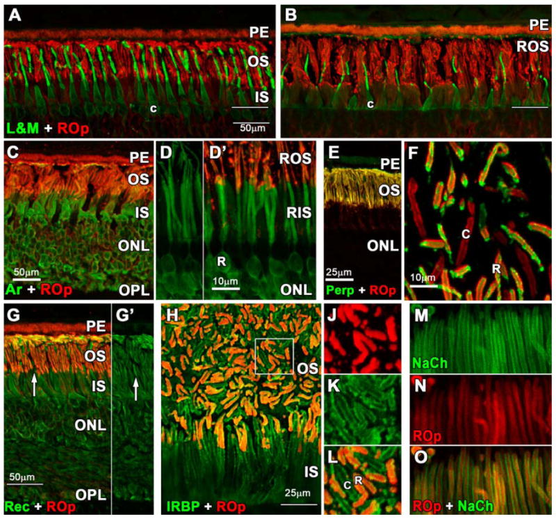
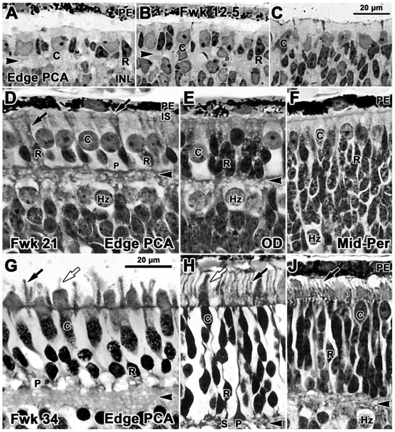
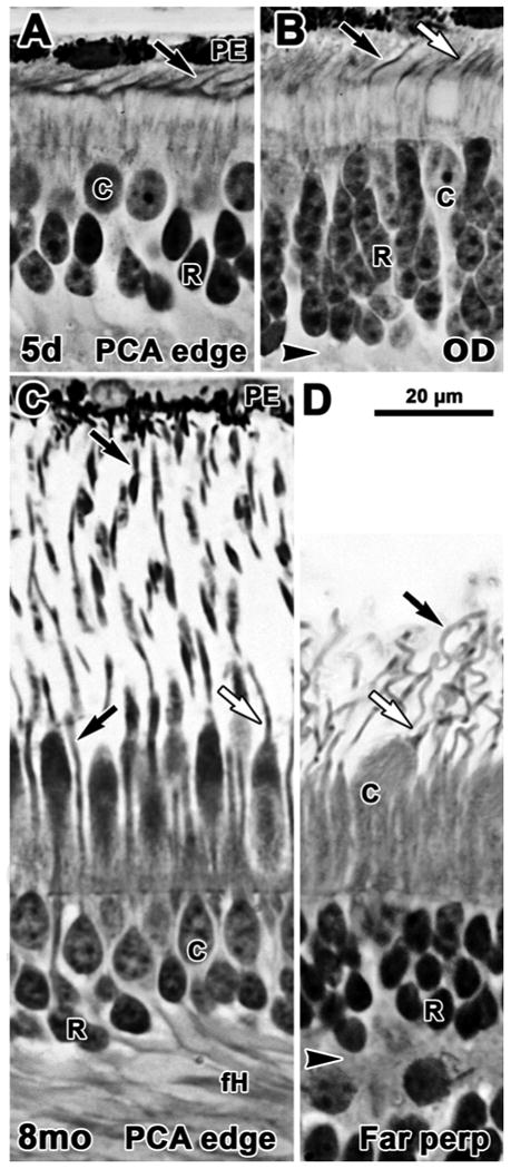
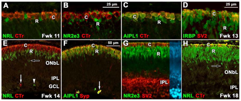
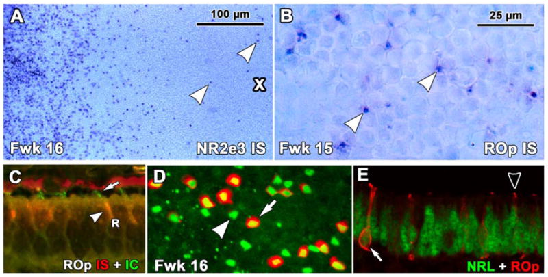
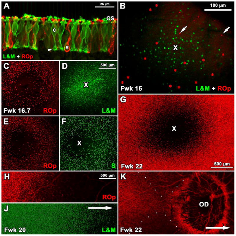
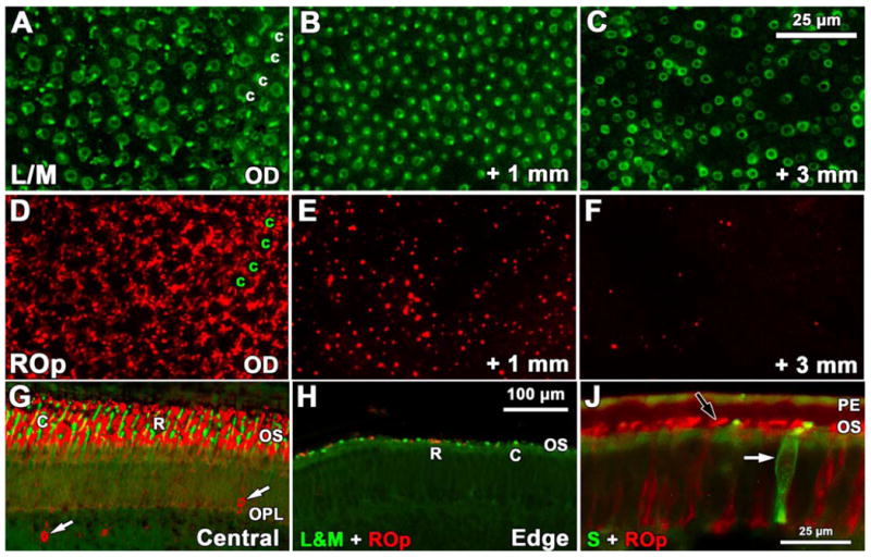
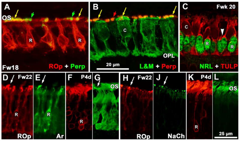
Human rods and cones are arranged in a precise spatial mosaic that is critical for optimal functioning of the visual system. However, the molecular processes that underpin specification of cell types within the mosaic are poorly understood. The progressive differentiation of human rods was tracked from fetal week (Fwk) 9 to postnatal (P) 8 months using immunocytochemical markers of key molecules that represent rod progression from post-mitotic precursors to outer segment-bearing functional photoreceptors. We find two phases associated with rod differentiation. The early phase begins in rods on the foveal edge at Fwk 10.5 when rods are first identified, and the rod-specific proteins NRL and NR2e3 are detected. By Fwk 11-12, these rods label for interphotoreceptor retinoid binding protein, recoverin, and aryl hydrocarbon receptor interacting protein-like 1. The second phase occurs over the next month with the appearance of rod opsin at Fwk 15, closely followed by the outer segment proteins rod GTP-gated sodium channel, rod arrestin, and peripherin. TULP is expressed relatively late at Fwk 18-20 in rods. Each phase proceeds across the retina in a central-peripheral order, such that rods in far peripheral retina are only entering the early phase at the same time that cells in central retina are entering their late phase. During the second half of gestation rods undergo an intracellular reorganization of these proteins, and cellular and OS elongation which continues into infancy. The progression of rod development shown here provides insight into the possible mechanisms underlying human retinal visual dysfunction when there are mutations affecting key rod-related molecules.
人类的视杆细胞和视锥细胞以精确的空间镶嵌模式排列,这对视觉系统的最佳功能至关重要。然而,对于这种镶嵌模式中细胞类型特化的分子过程,我们了解得还很少。利用代表视杆细胞从有丝分裂后前体到带有外段的功能性光感受器的关键分子的免疫细胞化学标记物,追踪了从胎儿第9周(Fwk)到出生后(P)8个月人类视杆细胞的逐步分化过程。我们发现视杆细胞分化有两个阶段。早期阶段始于Fwk 10.5时中央凹边缘的视杆细胞,此时首次识别出视杆细胞,并检测到视杆细胞特异性蛋白NRL和NR2e3。到Fwk 11 - 12时,这些视杆细胞标记为光感受器间类视黄醇结合蛋白、恢复蛋白和芳烃受体相互作用蛋白样1。第二阶段在下一个月出现,Fwk 15时出现视紫红质,紧接着是外段蛋白视杆细胞GTP门控钠通道、视杆细胞抑制蛋白和外周蛋白。TULP在视杆细胞中相对较晚在Fwk 18 - 20时表达。每个阶段都以中央到外周的顺序在整个视网膜上进行,使得远周边视网膜中的视杆细胞才刚刚进入早期阶段,而此时中央视网膜中的细胞正进入晚期阶段。在妊娠后半期,视杆细胞经历这些蛋白质的细胞内重组以及细胞和外段的伸长,这种过程会持续到婴儿期。这里所示的视杆细胞发育进程为当影响关键视杆细胞相关分子发生突变时人类视网膜视觉功能障碍的潜在机制提供了见解。