Getts Daniel R, Terry Rachael L, Getts Meghann Teague, Müller Marcus, Rana Sabita, Shrestha Bimmi, Radford Jane, Van Rooijen Nico, Campbell Iain L, King Nicholas J C
The Discipline of Pathology, School of Medical Sciences, The University of Sydney, Sydney NSW 2006, Australia.
J Exp Med. 2008 Sep 29;205(10):2319-37. doi: 10.1084/jem.20080421. Epub 2008 Sep 8.
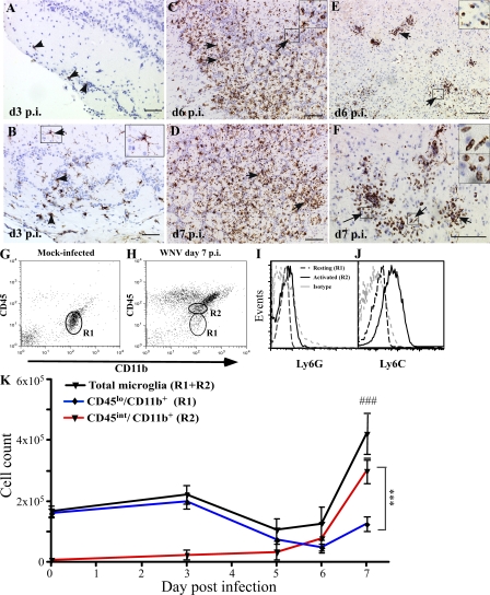
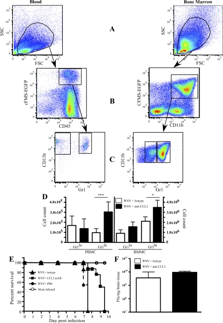
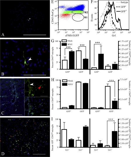
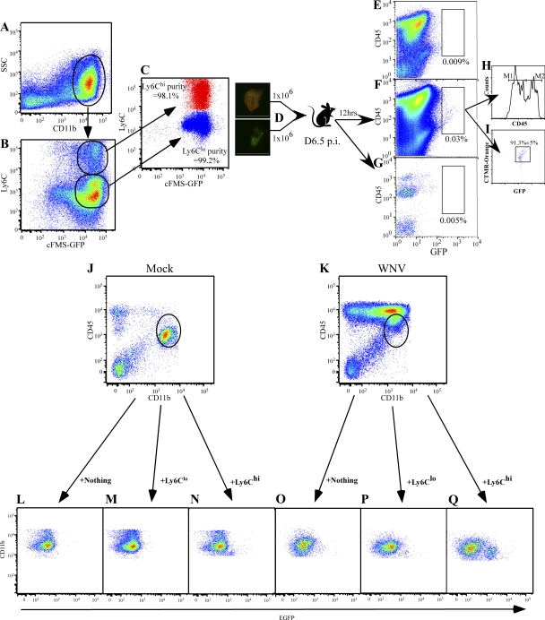
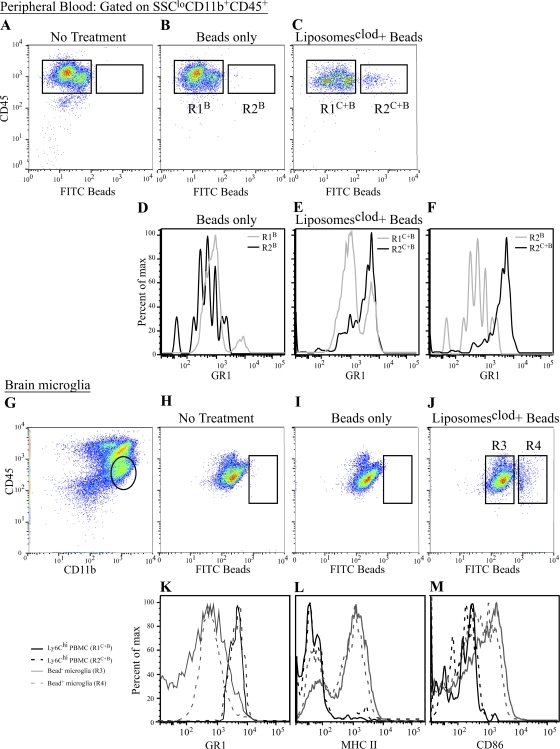
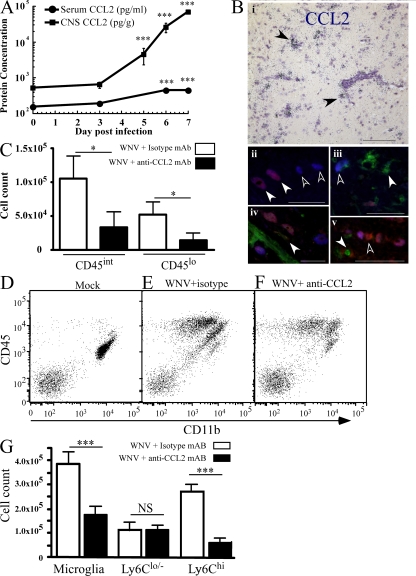
In a lethal West Nile virus (WNV) model, central nervous system infection triggered a threefold increase in CD45(int)/CD11b(+)/CD11c(-) microglia at days 6-7 postinfection (p.i.). Few microglia were proliferating, suggesting that the increased numbers were derived from a migratory precursor cell. Depletion of "circulating" (Gr1(-)(Ly6C(lo))CX3CR1(+)) and "inflammatory" (Gr1(hi)/Ly6C(hi)/CCR2(+)) classical monocytes during infection abrogated the increase in microglia. C57BL/6 chimeras reconstituted with cFMS-enhanced green fluorescent protein (EGFP) bone marrow (BM) showed large numbers of peripherally derived (GFP(+)) microglia expressing GR1(+)(Ly6C(+)) at day 7 p.i., suggesting that the inflammatory monocyte is a microglial precursor. This was confirmed by adoptive transfer of labeled BM (Ly6C(hi)/CD115(+)) or circulating inflammatory monocytes that trafficked to the WNV-infected brain and expressed a microglial phenotype. CCL2 is a chemokine that is highly expressed during WNV infection and important in inflammatory monocyte trafficking. Neutralization of CCL2 not only reduced the number of GFP(+) microglia in the brain during WNV infection but prolonged the life of infected animals. Therefore, CCL2-dependent inflammatory monocyte migration is critical for increases in microglia during WNV infection and may also play a pathogenic role during WNV encephalitis.
在致死性西尼罗河病毒(WNV)模型中,中枢神经系统感染在感染后(p.i.)第6 - 7天引发CD45(int)/CD11b(+)/CD11c(-)小胶质细胞数量增加三倍。很少有小胶质细胞增殖,这表明数量增加源自迁移前体细胞。感染期间“循环”(Gr1(-)(Ly6C(lo))CX3CR1(+))和“炎性”(Gr1(hi)/Ly6C(hi)/CCR2(+))经典单核细胞的耗竭消除了小胶质细胞的增加。用cFMS增强型绿色荧光蛋白(EGFP)骨髓(BM)重建的C57BL/6嵌合体在感染后第7天显示大量外周来源(GFP(+))的小胶质细胞表达GR1(+)(Ly6C(+)),这表明炎性单核细胞是小胶质细胞前体。通过将标记的骨髓(Ly6C(hi)/CD115(+))或循环炎性单核细胞过继转移到感染WNV的脑并表达小胶质细胞表型,这一点得到了证实。CCL2是一种趋化因子,在WNV感染期间高度表达且在炎性单核细胞迁移中起重要作用。CCL2的中和不仅减少了WNV感染期间脑中GFP(+)小胶质细胞的数量,还延长了感染动物的寿命。因此,CCL2依赖的炎性单核细胞迁移对于WNV感染期间小胶质细胞的增加至关重要,并且在WNV脑炎期间可能也起致病作用。