Chung Eun Jee, Lew Young Ju, Lee Hyo, Koh Hyoung Jun
Department of Ophthalmology, NHIC Ilsan Hospital, Gyounggi-do, Korea.
Korean J Ophthalmol. 2008 Sep;22(3):169-73. doi: 10.3341/kjo.2008.22.3.169.
To evaluate the usefulness of OCT retinal mapping in determining the configuration of a vitreomacular adhesion and selecting a meridian for entry into the subhyaloid space in patients with vitreomacular traction syndrome.
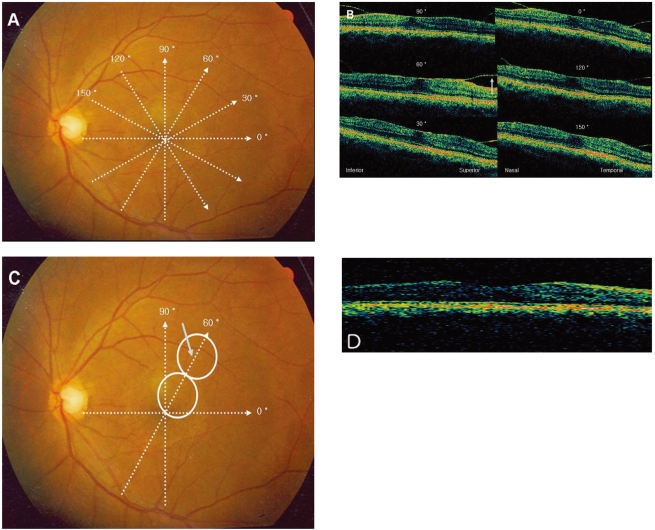
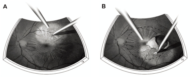
Six consecutive patients (6 eyes) with vitreomacular traction syndrome underwent vitrectomy with peeling of posterior hyaloid. Ocular coherence tomography (OCT) retinal mapping was performed preoperatively. Access to the subhyaloid space was made by creating an opening with a 25 gauge needle at a location where the detached posterior hyaloid membrane was farthest from the retinal surface. The location was selected based on six preoperative meridional OCT scans. The posterior hyaloid was then gently peeled off in a circular fashion around the fovea with a micropick. Visual acuity and foveal thicknesses were measured before the operation and 3 months afterwards.
After the operation, visual acuity improved and central macular thicknesses were reduced significantly in all six patients. The best corrected visual acuity improved from 0.4 to 0.75 with a mean increase by 3.5 lines on a Snellen chart 3 months after the operation. The mean foveal thickness was reduced from 406 micrometer to 241 micrometer. The restoration of foveal pit was observed in five patients. Neither intraoperative nor postoperative complications were observed during the follow up period.
An OCT retinal mapping program is a valuable diagnostic tool in understanding the configuration of vitreomacular adhesion and planning the surgical approach for operating on vitreomacular traction syndrome.
评估光学相干断层扫描(OCT)视网膜测绘在确定玻璃体黄斑粘连的形态以及为玻璃体黄斑牵引综合征患者选择进入玻璃体后间隙的子午线方面的实用性。
连续6例(6只眼)玻璃体黄斑牵引综合征患者接受了玻璃体后皮质剥除的玻璃体切除术。术前进行了OCT视网膜测绘。通过在脱离的玻璃体后皮质膜距视网膜表面最远的位置用25号针头制造一个开口进入玻璃体后间隙。该位置是根据术前6次子午线OCT扫描选定的。然后用微型剥膜器围绕黄斑以环形方式轻轻剥除玻璃体后皮质。在手术前和术后3个月测量视力和黄斑中心厚度。
术后,所有6例患者的视力均有提高,黄斑中心厚度显著降低。术后3个月,最佳矫正视力从0.4提高到0.75,在斯内伦视力表上平均提高3.5行。黄斑中心厚度平均从406微米降至241微米。5例患者观察到黄斑中心凹恢复。随访期间未观察到术中及术后并发症。
OCT视网膜测绘程序是了解玻璃体黄斑粘连形态和规划玻璃体黄斑牵引综合征手术方法的有价值的诊断工具。