Bühren Jens, Nagy Lana, Swanton Jennifer N, Kenner Shawn, MacRae Scott, Phipps Richard P, Huxlin Krystel R
University of Rochester Eye Institute, University of Rochester, Rochester, New York 14642, USA.
Invest Ophthalmol Vis Sci. 2009 Feb;50(2):634-43. doi: 10.1167/iovs.08-2277. Epub 2008 Oct 24.
To assess the contribution of corneal myofibroblasts to optical changes induced by photorefractive keratectomy (PRK) in a cat model.
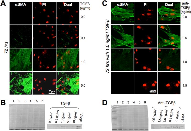
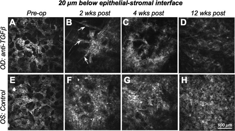
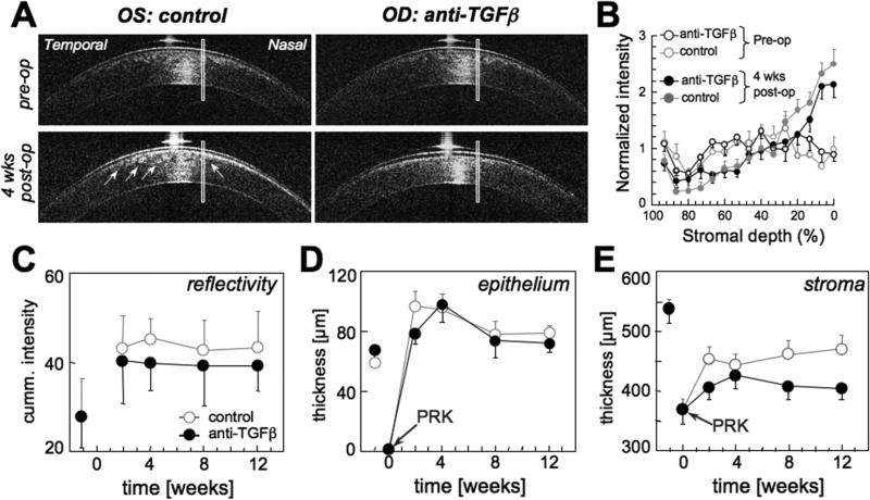
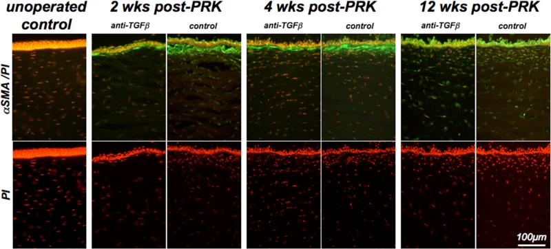
The transforming growth factor (TGF)-beta-dependence of feline corneal keratocyte differentiation into alpha-smooth muscle actin (alphaSMA)-positive myofibroblasts was first tested in vitro. Twenty-nine eyes of 16 cats were then treated with -10 D PRK in vivo and divided into two postoperative treatment groups that received either 100 microg anti-TGFbeta antibody for 7 days, followed by 50 microg dexamethasone for another 7 days to inhibit myofibroblast differentiation, or vehicle solution for 14 days (control eyes). Corneal thickness and reflectivity were measured by optical coherence tomography. Wavefront sensing was performed in the awake-behaving state before surgery and 2, 4, 8, and 12 weeks after surgery. Wound healing was monitored using in vivo confocal imaging and postmortem alphaSMA immunohistochemistry.
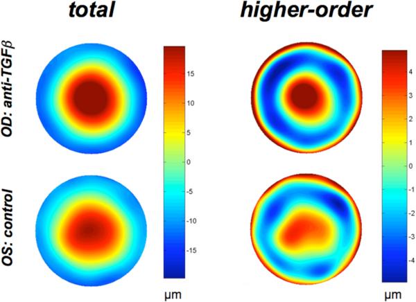
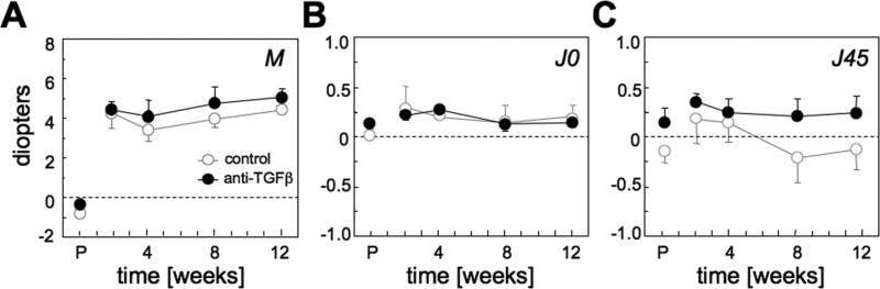
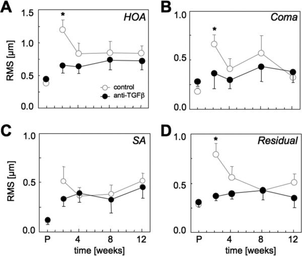
In culture, TGFbeta caused cat corneal keratocytes to differentiate into alphaSMA-positive myofibroblasts, an effect that was blocked by coincubation with anti-TGFbeta antibody. In vivo, anti-TGFbeta treatment after PRK resulted in less alphaSMA immunoreactivity in the subablation stroma, lower corneal reflectivity, less stromal regrowth, and lower nonspherical higher order aberration induction than in control eyes. However, there were no intergroup differences in epithelial regeneration or lower order aberration changes.
Anti-TGFbeta treatment reduced feline corneal myofibroblast differentiation in vitro and after PRK. It also decreased corneal haze and fine-grained irregularities in ocular wavefront after PRK, suggesting that attenuation of the differentiation of keratocytes into myofibroblast can significantly enhance optical quality after refractive surface ablations.
在猫模型中评估角膜肌成纤维细胞对光性屈光性角膜切削术(PRK)诱导的光学变化的作用。
首先在体外测试猫角膜基质细胞分化为α-平滑肌肌动蛋白(αSMA)阳性肌成纤维细胞对转化生长因子(TGF)-β的依赖性。然后对16只猫的29只眼进行体内-10 D的PRK治疗,并分为两个术后治疗组,一组接受100μg抗TGFβ抗体治疗7天,随后接受50μg地塞米松再治疗7天以抑制肌成纤维细胞分化,另一组接受赋形剂溶液治疗14天(对照眼)。通过光学相干断层扫描测量角膜厚度和反射率。在手术前以及手术后2、4、8和12周在清醒行为状态下进行波前传感。使用体内共聚焦成像和死后αSMA免疫组织化学监测伤口愈合。
在培养中,TGFβ使猫角膜基质细胞分化为αSMA阳性肌成纤维细胞,与抗TGFβ抗体共同孵育可阻断该效应。在体内,PRK后抗TGFβ治疗导致消融下基质中的αSMA免疫反应性降低、角膜反射率降低、基质再生减少以及非球面高阶像差诱导低于对照眼。然而,两组在上皮再生或低阶像差变化方面没有差异。
抗TGFβ治疗在体外和PRK后均可减少猫角膜肌成纤维细胞分化。它还减少了PRK后角膜混浊和眼波前的细颗粒不规则性,表明基质细胞向肌成纤维细胞分化的减弱可显著提高屈光表面消融后的光学质量。