Guimaraes Alexander R, Rakhlin Elena, Weissleder Ralph, Thayer Sarah P
Department of Radiology, Center for Molecular Imaging Research, Massachusetts General Hospital, Charlestown, MA, USA.
Pancreas. 2008 Nov;37(4):440-4. doi: 10.1097/MPA.0b013e31817c5113.
The sonic hedgehog (Shh) pathway has an established role in pancreatic cancer (pancreatic adenocarcinoma [PDAC]). We tested whether magnetic resonance imaging measures of vascular volume fraction (VVF) using magnetic iron oxide nanoparticles are sensitive to the antiangiogenic effect of targeted Shh therapies in a PDAC xenograft model.
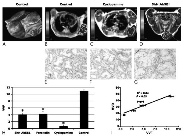
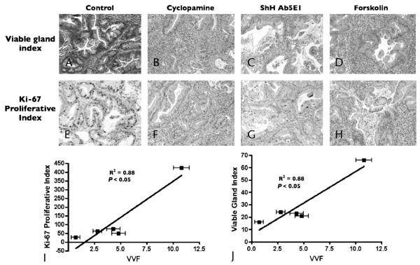
Pancreatic adenocarcinoma xenograft lines were subcutaneously implanted into nude mice (n = 19 samples within 4 groups). Therapies were targeted to 3 loci of the Shh signaling pathway (anti-Shh antibody, cyclopamine, or forskolin). Magnetic resonance imaging (4.7-T Bruker Pharmascan) was performed (after 1 week of intraperitoneal therapy) before and after intravenous injection of MION-47. Vascular volume fraction was quantified as DeltaR2 (from multicontrast T2 sequences) and normalized to an assumed VVF in muscle of 3%. Linear regression compared VVF to histological indices including microvessel density (MVD), viable gland density (VGD), and proliferative index (PI).
In response to anti-Hh treatment, tumors showed a decrease in VGD, PI, MVD, and VVF compared with controls (P < 0.001). Vascular volume fraction was compared with histological indicators of response: PI (R2 = 0.88; P < 0.05), VGD (R2 = 0.87; P< 0.05), and MVD (R2 = 0.85; P < 0.05).
Magnetic resonance imaging VVF using magnetic iron oxide nanoparticles may serve as a noninvasive measure of biological response to Shh PDAC therapy with easy translation to the clinic.
音猬因子(Shh)信号通路在胰腺癌(胰腺腺癌[PDAC])中已明确发挥作用。我们测试了使用磁性氧化铁纳米颗粒进行磁共振成像测量血管容积分数(VVF),是否对PDAC异种移植模型中靶向Shh治疗的抗血管生成作用敏感。
将胰腺腺癌异种移植系皮下植入裸鼠体内(4组共19个样本)。治疗针对Shh信号通路的3个位点(抗Shh抗体、环杷明或福司可林)。在腹腔内治疗1周后,静脉注射MION - 47前后进行磁共振成像(4.7 - T布鲁克药物扫描仪)。血管容积分数定量为ΔR2(来自多对比度T2序列),并归一化为假定肌肉中的VVF为3%。线性回归将VVF与包括微血管密度(MVD)、存活腺密度(VGD)和增殖指数(PI)在内的组织学指标进行比较。
与对照组相比,抗Hh治疗后肿瘤的VGD、PI、MVD和VVF均降低(P < 0.001)。将血管容积分数与反应的组织学指标进行比较:PI(R2 = 0.88;P < 0.05)、VGD(R2 = 0.87;P < 0.05)和MVD(R2 = 0.85;P < 0.05)。
使用磁性氧化铁纳米颗粒的磁共振成像VVF可作为对Shh - PDAC治疗生物反应的非侵入性测量方法,易于转化到临床应用。