Kampmann Andreas, Fernández Borja, Deindl Elisabeth, Kubin Thomas, Pipp Frederic, Eitenmüller Inka, Hoefer Imo E, Schaper Wolfgang, Zimmermann René
Research Group Vascular Genomics, Kerckhoff Clinic, Bad Nauheim, Germany.
Mol Cell Biochem. 2009 Feb;322(1-2):15-23. doi: 10.1007/s11010-008-9935-x. Epub 2008 Nov 4.
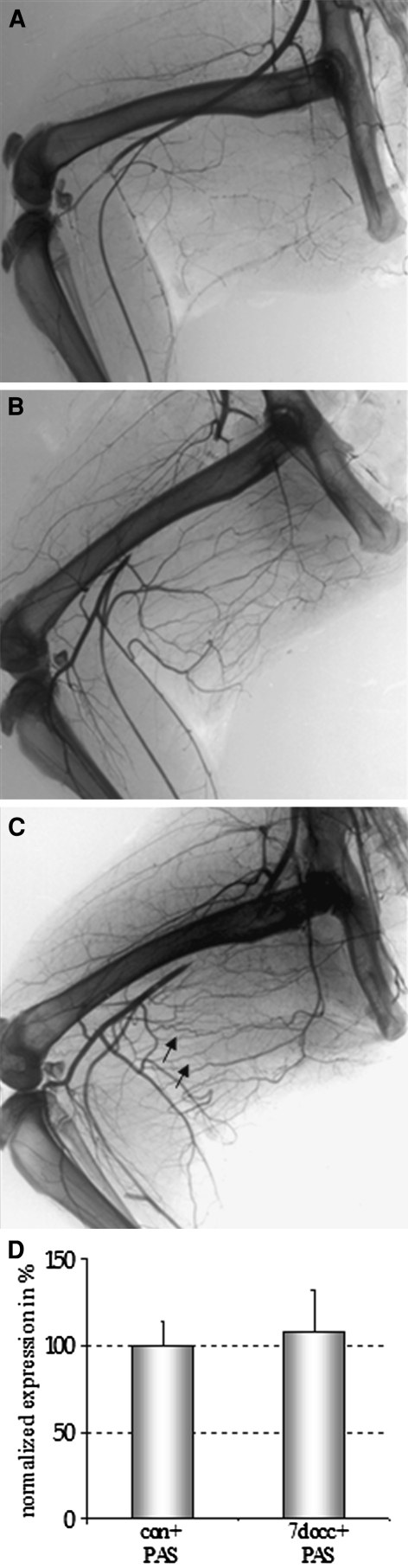
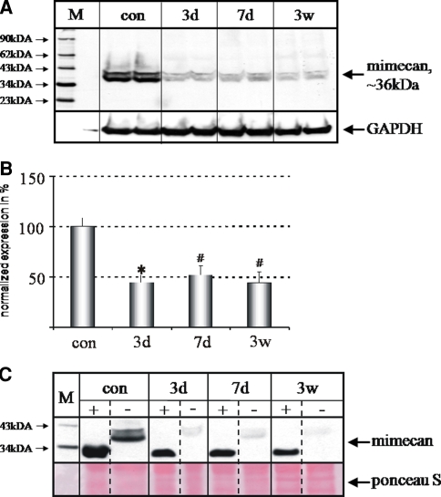
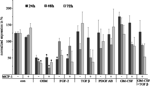
Arteriogenesis or collateral growth is able to compensate for the stenosis of major arteries. Using differential display RT-PCR on growing and quiescent collateral arteries in a rabbit femoral artery ligation model, we cloned the rabbit full-length cDNA of osteoglycin/mimecan. Osteoglycin was present in the adventitia of collateral arteries as a glycosylated protein without keratan sulfate side chains, mainly produced by smooth muscle cells (SMCs) and perivascular fibroblasts. Northern blot, Western blot, and immunohistochemistry confirmed a collateral artery-specific downregulation of osteoglycin from 6 h to 3 weeks after the onset of arteriogenesis. Treatment of primary SMCs with the arteriogenic protein fibroblast growth factor-2 (FGF-2) resulted in a similar reduction of osteoglycin expression as observed in vivo. Application of the FGF-2 inhibitor polyanethole sulfonic acid (PAS) blocked the downregulation of osteoglycin and interfered with arteriogenesis. From our study we conclude that downregulation of osteoglycin is a fundamental requirement for proper arteriogenesis.
动脉生成或侧支生长能够补偿主要动脉的狭窄。在兔股动脉结扎模型中,利用差异显示逆转录聚合酶链反应(RT-PCR)对生长中的和静止的侧支动脉进行研究,我们克隆了骨形成蛋白/ mimecan的兔全长cDNA。骨形成蛋白作为一种无硫酸角质素侧链的糖基化蛋白存在于侧支动脉的外膜中,主要由平滑肌细胞(SMC)和血管周成纤维细胞产生。Northern印迹、Western印迹和免疫组织化学证实,在动脉生成开始后的6小时至3周内,骨形成蛋白在侧支动脉中特异性下调。用动脉生成蛋白成纤维细胞生长因子-2(FGF-2)处理原代SMC,导致骨形成蛋白表达出现与体内观察到的类似降低。应用FGF-2抑制剂聚茴香脑磺酸(PAS)可阻断骨形成蛋白的下调,并干扰动脉生成。从我们的研究中我们得出结论,骨形成蛋白的下调是正常动脉生成的基本要求。