de Diego Carlos, Pai Rakesh K, Chen Fuhua, Xie Lai-Hua, De Leeuw Jan, Weiss James N, Valderrábano Miguel
UCLA Cardiovascular Research Laboratory, Department of Medicine, Division of Cardiology, David Geffen School of Medicine at UCLA, Los Angeles, CA 90095, USA.
Circulation. 2008 Dec 2;118(23):2330-7. doi: 10.1161/CIRCULATIONAHA.108.789149. Epub 2008 Nov 17.
Electrophysiological changes promoting arrhythmias during acute regional ischemia/reperfusion are challenging to study in intact cardiac tissue because of complex 3-dimensional myocardial and vascular geometry. We characterized electrophysiological alterations and arrhythmias during regional ischemia/reperfusion in a simpler 2-dimensional geometry of cultured neonatal rat ventricular myocyte monolayers.
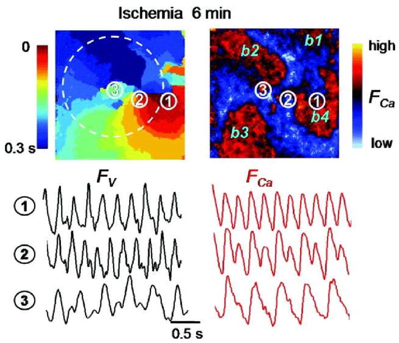
Optical mapping of intracellular Ca (Ca(i)) and voltage was performed with the use of Rhod 2-AM and Rh-237, respectively. Regional ischemia was mimicked by covering the central portion of monolayer with a glass coverslip, and reperfusion was mimicked by removing the coverslip. Monolayers were stained with fluorescent antibodies to detect total and dephosphorylated connexin-43 at various time points. During coverslip ischemia, action potential duration shortened, Ca(i) transient duration was prolonged, and local conduction velocity (CV) slowed progressively, with loss of excitability after 10.6 +/- 3.6 minutes. CV slowing was accompanied by connexin-43 dephosphorylation. During ischemia, spontaneous reentry occurred in 5 of 11 monolayers, initiated by extrasystoles arising from the border zone or unidirectional conduction block of paced beats. On reperfusion, excitability recovered within 1.0 +/- 0.8 minutes, but CV remained depressed for 9.0 +/- 3.0 minutes, promoting reentry in the reperfused zone. As connexin-43 phosphorylation recovered in the reperfused zone, CV normalized, and arrhythmias resolved.
Acute regional ischemia/reperfusion in neonatal rat ventricular myocyte monolayers recapitulates electrophysiological alterations and arrhythmias similar to those observed during acute coronary occlusion/reperfusion in intact hearts. During early reperfusion, slow recovery from connexin-43 dephosphorylation leads to persistent CV slowing, creating a highly arrhythmogenic substrate.
由于心肌和血管复杂的三维几何结构,在完整心脏组织中研究急性局部缺血/再灌注期间促进心律失常的电生理变化具有挑战性。我们在培养的新生大鼠心室肌细胞单层的更简单二维几何结构中,对局部缺血/再灌注期间的电生理改变和心律失常进行了特征描述。
分别使用Rhod 2-AM和Rh-237对细胞内钙(Ca(i))和电压进行光学映射。用玻璃盖玻片覆盖单层的中央部分模拟局部缺血,移除盖玻片模拟再灌注。在不同时间点用荧光抗体对单层进行染色,以检测总连接蛋白43和去磷酸化连接蛋白43。在盖玻片缺血期间,动作电位时程缩短,Ca(i)瞬变时程延长,局部传导速度(CV)逐渐减慢,在10.6±3.6分钟后兴奋性丧失。CV减慢伴随着连接蛋白43的去磷酸化。在缺血期间,11个单层中有5个出现自发折返,由边界区的期外收缩或起搏搏动的单向传导阻滞引发。再灌注时,兴奋性在1.0±0.8分钟内恢复,但CV在9.0±3.0分钟内仍受抑制,促进了再灌注区的折返。随着再灌注区连接蛋白43磷酸化的恢复,CV恢复正常,心律失常消失。
新生大鼠心室肌细胞单层的急性局部缺血/再灌注概括了与完整心脏急性冠状动脉闭塞/再灌注期间观察到的相似的电生理改变和心律失常。在早期再灌注期间,连接蛋白43去磷酸化的缓慢恢复导致CV持续减慢,形成高度致心律失常的基质。