Meyers Philip M, Schumacher H Christian, Gray William A, Fifi Johanna, Gaudet John G, Heyer Eric J, Chong Ji Y
Department of Radiology, Columbia University, College of Physicians & Surgeons, Neurological Institute of New York, New York, New York 10032, USA.
J Neuroimaging. 2009 Jul;19(3):266-70. doi: 10.1111/j.1552-6569.2008.00278.x. Epub 2008 Oct 24.
Intracranial artery stenosis is assumed to represent atherosclerotic plaque. Catheter cerebral arteriography shows that intracranial stenosis may progress, regress, or remain unchanged. It is counterintuitive that atherosclerotic plaque should spontaneously regress, raising questions about the composition of intracranial stenoses. Little is known about this disease entity in vivo. We provide the first demonstration of in vivo atherosclerotic plaque with intraplaque hemorrhage using intravascular ultrasound (IVUS).
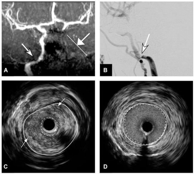
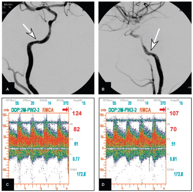
A 35-year-old man with multiple vascular risk factors presented with recurrent stroke failing medical therapy. Imaging demonstrated left internal carotid artery occlusion, severe intracranial right internal carotid artery stenosis, and cerebral perfusion failure. Cerebral arteriography with IVUS confirmed 85% stenosis of the petrous right carotid artery due to atherosclerotic plaque with intraplaque hemorrhage. Intracranial stent-supported angioplasty was performed with IRB approval. The patient recovered without complication.
This case supports the premise that symptomatic intracranial stenosis can be caused by atherosclerotic plaque complicated by intraplaque hemorrhage similar to coronary artery plaque. IVUS provides additional characteristics that define intracranial atherosclerosis and high-risk features. To our knowledge, this is the first report of stroke due to unstable atherosclerotic plaque with intraplaque hemorrhage in vivo.
颅内动脉狭窄被认为代表动脉粥样硬化斑块。导管脑血管造影显示颅内狭窄可能进展、消退或保持不变。动脉粥样硬化斑块会自发消退,这有悖于直觉,引发了关于颅内狭窄成分的疑问。关于这种疾病实体在体内的情况知之甚少。我们首次使用血管内超声(IVUS)在体内证实了伴有斑块内出血的动脉粥样硬化斑块。
一名35岁有多种血管危险因素的男性,因药物治疗无效出现复发性中风。影像学检查显示左侧颈内动脉闭塞、右侧颅内颈内动脉严重狭窄以及脑灌注衰竭。脑血管造影联合IVUS证实右侧岩部颈动脉因伴有斑块内出血的动脉粥样硬化斑块而出现85%的狭窄。在获得机构审查委员会批准后进行了颅内支架辅助血管成形术。患者康复且无并发症。
本病例支持以下前提,即有症状的颅内狭窄可能由类似于冠状动脉斑块的伴有斑块内出血的动脉粥样硬化斑块引起。IVUS提供了定义颅内动脉粥样硬化和高危特征的额外特征。据我们所知,这是第一例关于体内不稳定动脉粥样硬化斑块伴有斑块内出血导致中风的报告。