Pegoraro-Krook Maria Inês, Dutka-Souza Jeniffer de Cassia Rillo, Marino Viviane Cristina de Castro
Department of Speech Pathology and Audiology, Bauru School of Dentistry and Hospital for Rehabilitation of Craniofacial Anomalies, University of São Paulo, Bauru, SP, Brazil.
J Appl Oral Sci. 2008 May-Jun;16(3):181-8. doi: 10.1590/s1678-77572008000300004.
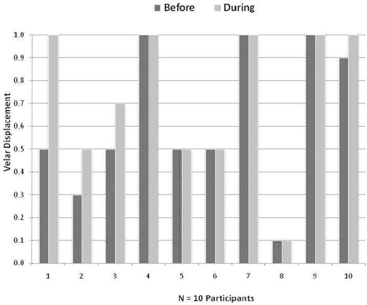
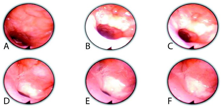
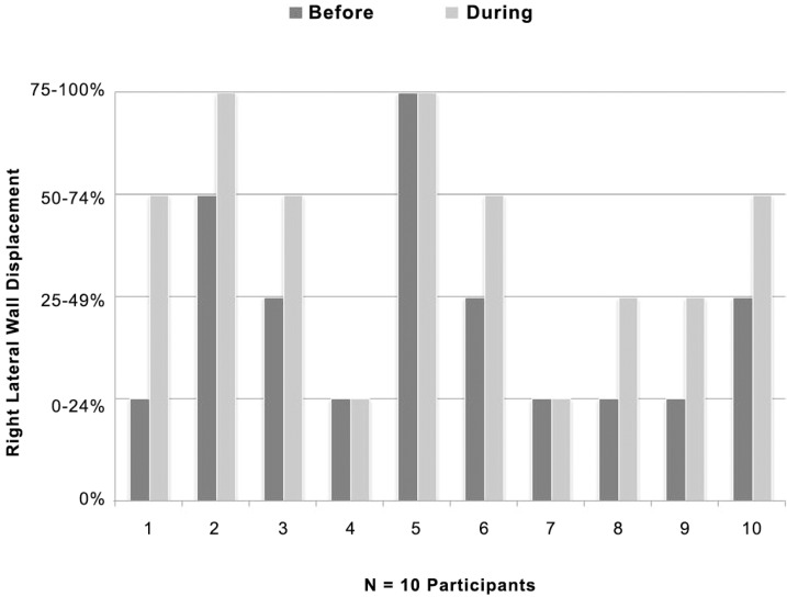
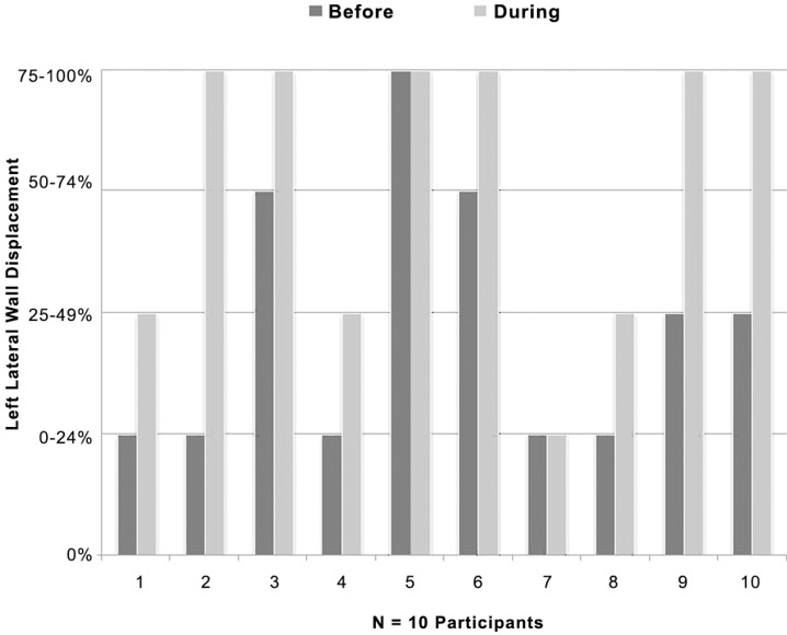
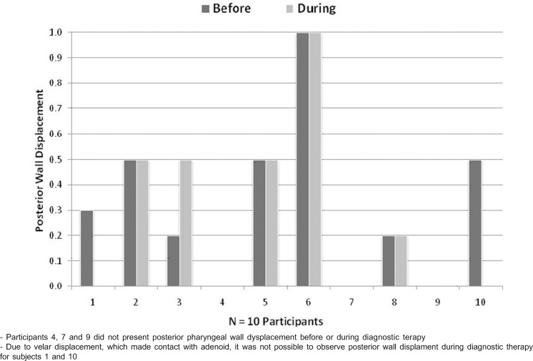
Nasoendoscopy is an important tool for assessing velopharyngeal function. The purpose of this study was to analyze velar and pharyngeal wall movement and velopharyngeal gap during nasoendoscopic evaluation of the velopharynx before and during diagnostic therapy. Nasoendoscopic recordings of 10 children with operated cleft lip and palate were analyzed according to the International Working Group Guidelines. Ratings of movement of velum and pharyngeal walls, and size, location and shape of gaps were analyzed by 3 speech-language pathologists (SLPs). Imaging was obtained during repetitions of the syllable /pa/ during a single nasoendoscopic evaluation: (a) before diagnostic therapy, and (b) after the children were instructed to impound and increase intraoral air pressure (diagnostic therapy). Once the patients impounded and directed air pressure orally, the displacement of the velum, right, left and posterior pharyngeal walls increased 40, 70, 80, and 10%, respectively. Statistical significance for displacement was found only for right and left lateral pharyngeal walls. Reduction in gap size was observed for 30% of the patients and other 40% of the gaps disappeared. Changes in gap size were found to be statistically significant between the two conditions. In nasoendoscopic assessment, the full potential of velopharyngeal displacement may not be completely elicited when the patient is asked only to repeat a speech stimulus. Optimization of information can be done with the use of diagnostic therapy's strategies to manipulate VP function. Assuring the participation of the SLP to conduct diagnostic therapy is essential for management of velopharyngeal dysfunction.
鼻内镜检查是评估腭咽功能的重要工具。本研究的目的是分析在诊断性治疗前后对腭咽进行鼻内镜评估时软腭和咽壁的运动以及腭咽间隙。根据国际工作组指南,对10例唇腭裂手术患儿的鼻内镜记录进行了分析。3名言语语言病理学家(SLP)分析了软腭和咽壁的运动评级以及间隙的大小、位置和形状。在单次鼻内镜评估中重复发/pa/音节时进行成像:(a)诊断性治疗前,以及(b)在指导患儿紧闭并增加口腔内气压(诊断性治疗)后。一旦患者紧闭并引导口腔内气压,软腭、右侧、左侧和咽后壁的位移分别增加了40%、70%、80%和10%。仅在右侧和左侧咽壁发现位移具有统计学意义。30%的患者间隙尺寸减小,另外40%的间隙消失。两种情况下间隙尺寸的变化具有统计学意义。在鼻内镜评估中,当仅要求患者重复语音刺激时,腭咽位移的全部潜力可能无法完全激发。利用诊断性治疗策略来操纵腭咽功能可以优化信息。确保言语语言病理学家参与进行诊断性治疗对于腭咽功能障碍的管理至关重要。