Carr Amanda-Jayne, Vugler Anthony, Lawrence Jean, Chen Li Li, Ahmado Ahmed, Chen Fred K, Semo Ma'ayan, Gias Carlos, da Cruz Lyndon, Moore Harry D, Walsh James, Coffey Peter J
Institute of Ophthalmology, University College London, London, UK.
Mol Vis. 2009;15:283-95. Epub 2009 Feb 6.
To examine the ability of retinal pigment epithelial (RPE) cells derived from human embryonic stem cells (HESC) to phagocytose photoreceptor outer segments, and to determine whether exposure to human retina induces any morphological changes in these cells.
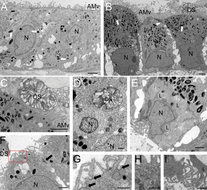
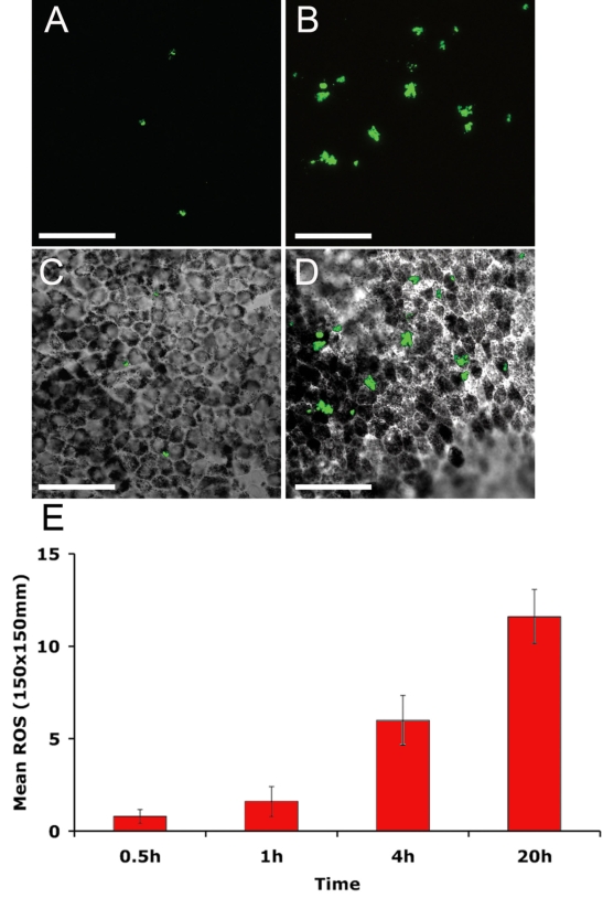
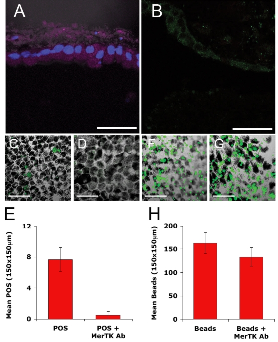
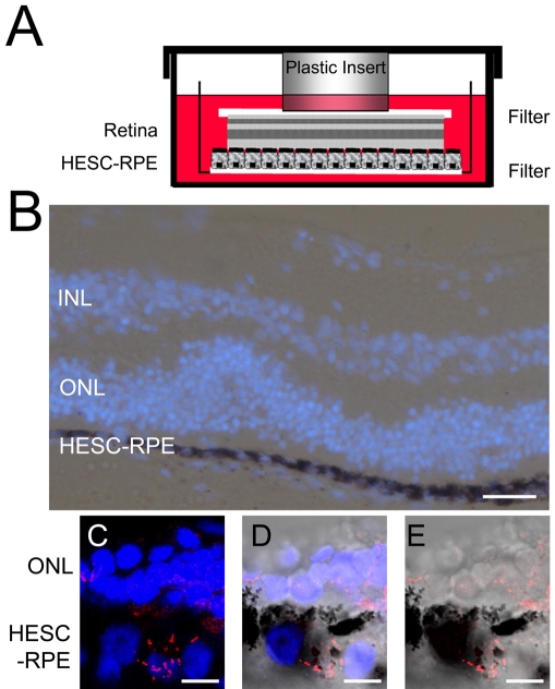
HESC-RPE cells were derived from a super-confluent preparation of the Shef1 HESC line. Pigmented colonies were isolated and expanded into pigmented monolayers on Matrigel matrix-coated dishes or filters. Cells were exposed to fluorescently labeled outer segments isolated from the porcine eye and assessed for phagocytic activity at regular intervals. Expression of molecules associated with RPE phagocytosis was analyzed by RT-PCR, immunocytochemistry, and western blot. The role of Mer Tyrosine Kinase (MERTK) in the phagocytosis of outer segments was investigated using antibodies directed against MERTK to block function. In a novel approach, cells were also exposed to fresh human neural retina tissue then examined by electron microscopy for evidence of phagocytosis and changes in cell morphology.
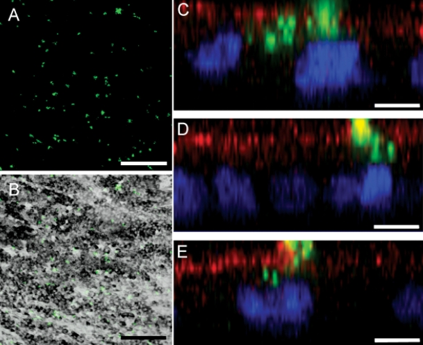
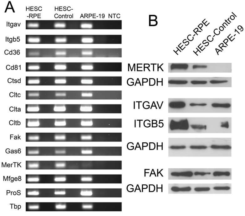
HESC-derived RPE cells are capable of phagocytosing isolated porcine outer segments and express molecules associated with RPE-specific phagocytosis, including MERTK. Pre-incubation with antibodies against MERTK blocked phagocytosis of photoreceptor outer segments, but not polystyrene beads. HESC-RPE cells also phagocytosed outer segments in a novel human retinal explant system. Furthermore co-culture adjacent to human retina tissue in this preparation resulted in the appearance of features in HESC-derived RPE cells normally observed only as the RPE matures.
The ingestion of photoreceptor outer segments from an isolated population and an artificial ex vivo human retina system demonstrates HESC-derived RPE cells are functional. HESC-derived RPE possess the relevant molecules required for phagocytosis, including MERTK, which is essential for the phagocytosis of outer segments but not latex beads. Furthermore, some changes observed in cell morphology after co-culture with human retina may have implications for understanding the full development and differentiation of RPE cells.
研究源自人类胚胎干细胞(HESC)的视网膜色素上皮(RPE)细胞吞噬光感受器外节的能力,并确定暴露于人类视网膜是否会诱导这些细胞发生任何形态学变化。
HESC-RPE细胞源自Shef1 HESC系的超汇合培养物。分离出色素沉着的集落,并在基质胶包被的培养皿或滤膜上扩增为色素沉着的单层细胞。将细胞暴露于从猪眼中分离出的荧光标记外节,并定期评估吞噬活性。通过逆转录聚合酶链反应(RT-PCR)、免疫细胞化学和蛋白质免疫印迹分析与RPE吞噬相关分子的表达。使用针对Mer酪氨酸激酶(MERTK)的抗体阻断其功能,研究MERTK在光感受器外节吞噬中的作用。采用一种新方法,将细胞也暴露于新鲜的人类神经视网膜组织,然后通过电子显微镜检查吞噬作用和细胞形态变化的证据。
源自HESC的RPE细胞能够吞噬分离的猪外节,并表达与RPE特异性吞噬相关的分子,包括MERTK。用抗MERTK抗体预孵育可阻断光感受器外节的吞噬作用,但不影响聚苯乙烯珠的吞噬。HESC-RPE细胞在一种新的人类视网膜外植体系统中也能吞噬外节。此外,在此制备物中与人类视网膜组织共培养相邻区域,导致源自HESC的RPE细胞出现通常仅在RPE成熟时才观察到的特征。
从分离群体和人工离体人类视网膜系统中摄取光感受器外节表明源自HESC的RPE细胞具有功能。源自HESC的RPE细胞具有吞噬所需的相关分子,包括MERTK,它对光感受器外节的吞噬至关重要,但对乳胶珠的吞噬不是必需的。此外,与人类视网膜共培养后在细胞形态上观察到的一些变化可能对理解RPE细胞的完全发育和分化具有启示意义。