Molnar Joseph A, Lew Wesley K, Rapp Derek A, Gordon E Stanley, Voignier Denise, Rushing Scott, Willner William
Department of Plastic and Reconstructive Surgery, Wake Forest University School of Medicine, Winston-Salem, NC, USA.
Eplasty. 2009;9:e4. Epub 2009 Jan 12.
We developed a Web-based, blinded, prospective, randomized, multicenter trial, using standardized digital photography to clinically evaluate hand burn depth and accurately determine wound area with digital planimetry.
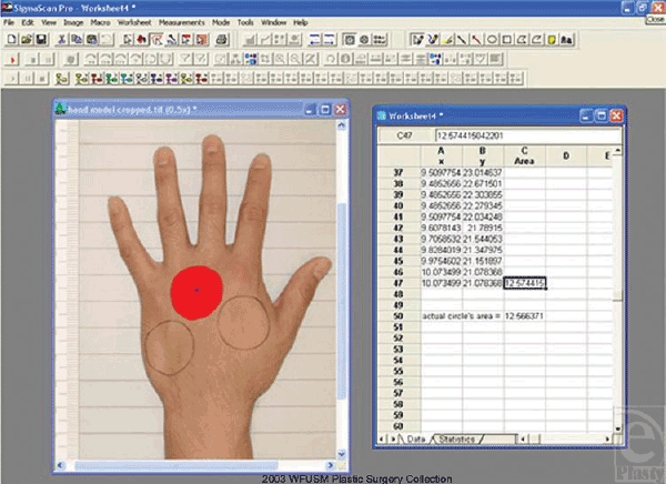
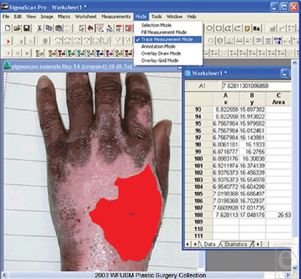
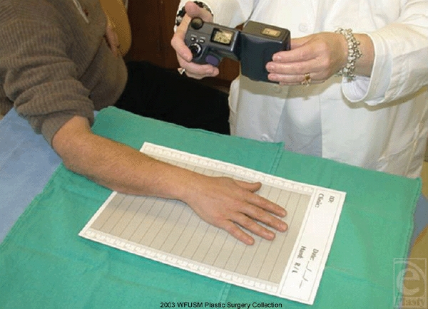
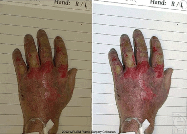
Photos in each center were taken with identical digital cameras with standardized settings on a custom backdrop developed at Wake Forest University containing a gray, white, black, and centimeter scale. The images were downloaded, transferred via the Web, and stored on servers at the principal investigator's home institution. Color adjustments to each photo were made using Adobe Photoshop 6.0 (Adobe, San Jose, Calif). In an initial pilot study, model hands marked with circles of known areas were used to determine the accuracy of the planimetry technique. Two-dimensional digital planimetry using SigmaScan Pro 5.0 (SPSS Science, Chicago, Ill) was used to calculate wound area from the digital images.
Digital photography is a simple and cost-effective method for quantifying wound size when used in conjunction with digital planimetry (SigmaScan) and photo enhancement (Adobe Photoshop) programs. The accuracy of the SigmaScan program in calculating predetermined areas was within 4.7% (95% CI, 3.4%-5.9%). Dorsal hand burns of the initial 20 patients in a national study involving several centers were evaluated with this technique. Images obtained by individuals denying experience in photography proved reliable and useful for clinical evaluation and quantification of wound area.
Standardized digital photography may be used quantitatively in a Web-based, multicenter trial of burn care. This technique could be modified for other medical studies with visual endpoints.
我们开展了一项基于网络的、盲法、前瞻性、随机、多中心试验,使用标准化数码摄影对烧伤深度进行临床评估,并通过数字平面测量法准确测定伤口面积。
每个中心使用相同的数码相机,在维克森林大学开发的定制背景布上进行拍摄,背景布上有灰色、白色、黑色和厘米刻度尺,相机设置为标准化设置。图像被下载,通过网络传输,并存储在主要研究者所在机构的服务器上。使用Adobe Photoshop 6.0(Adobe公司,加利福尼亚州圣何塞)对每张照片进行色彩调整。在初步的试点研究中,使用标有已知面积圆圈的模型手来确定平面测量技术的准确性。使用SigmaScan Pro 5.0(SPSS Science公司,伊利诺伊州芝加哥)进行二维数字平面测量,从数字图像中计算伤口面积。
数码摄影与数字平面测量(SigmaScan)和照片增强(Adobe Photoshop)程序结合使用时,是一种简单且经济高效的伤口大小量化方法。SigmaScan程序计算预定面积的准确性在4.7%以内(95%可信区间,3.4%-5.9%)。在一项涉及多个中心的全国性研究中,使用该技术对最初20例患者的手背烧伤进行了评估。由不具备摄影经验的人员获取的图像被证明对伤口面积的临床评估和量化可靠且有用。
标准化数码摄影可在基于网络的烧伤护理多中心试验中进行定量使用。该技术可针对其他具有视觉终点的医学研究进行改进。