Zhang Dan, Shooshtarizadeh Peiman, Laventie Benoît-Joseph, Colin Didier André, Chich Jean-François, Vidic Jasmina, de Barry Jean, Chasserot-Golaz Sylvette, Delalande François, Van Dorsselaer Alain, Schneider Francis, Helle Karen, Aunis Dominique, Prévost Gilles, Metz-Boutigue Marie-Hélène
INSERM U575, Physiopathologie du Système Nerveux, Strasbourg, France.
PLoS One. 2009;4(2):e4501. doi: 10.1371/journal.pone.0004501. Epub 2009 Feb 19.
Antimicrobial peptides derived from the natural processing of chromogranin A (CgA) are co-secreted with catecholamines upon stimulation of chromaffin cells. Since PMNs play a central role in innate immunity, we examine responses by PMNs following stimulation by two antimicrobial CgA-derived peptides.
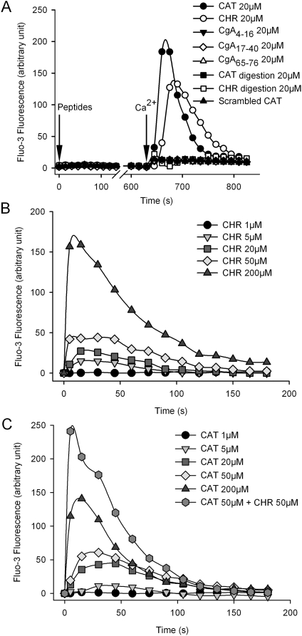
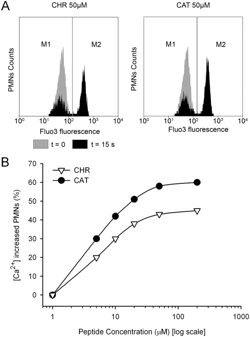
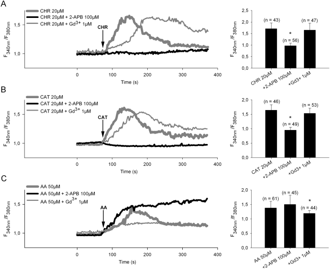
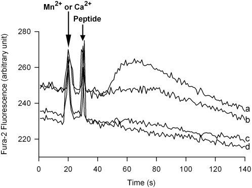
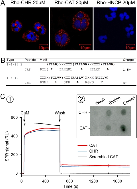
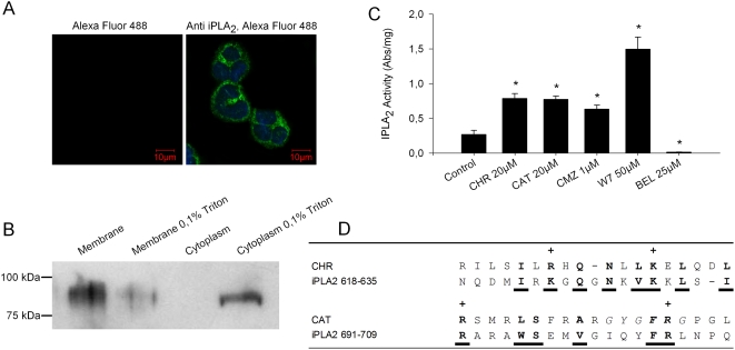
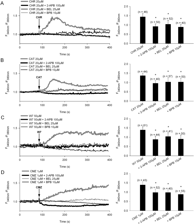
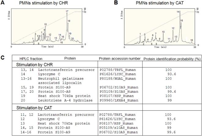
METHODOLOGY/PRINCIPAL FINDINGS: PMNs were treated with different concentrations of CgA-derived peptides in presence of several drugs. Calcium mobilization was observed by using flow cytometry and calcium imaging experiments. Immunocytochemistry and confocal microscopy have shown the intracellular localization of the peptides. The calmodulin-binding and iPLA2 activating properties of the peptides were shown by Surface Plasmon Resonance and iPLA2 activity assays. Finally, a proteomic analysis of the material released after PMNs treatment with CgA-derived peptides was performed by using HPLC and Nano-LC MS-MS. By using flow cytometry we first observed that after 15 s, in presence of extracellular calcium, Chromofungin (CHR) or Catestatin (CAT) induce a concentration-dependent transient increase of intracellular calcium. In contrast, in absence of extra cellular calcium the peptides are unable to induce calcium depletion from the stores after 10 minutes exposure. Treatment with 2-APB (2-aminoethoxydiphenyl borate), a store operated channels (SOCs) blocker, inhibits completely the calcium entry, as shown by calcium imaging. We also showed that they activate iPLA2 as the two CaM-binding factors (W7 and CMZ) and that the two sequences can be aligned with the two CaM-binding domains reported for iPLA2. We finally analyzed by HPLC and Nano-LC MS-MS the material released by PMNs following stimulation by CHR and CAT. We characterized several factors important for inflammation and innate immunity.
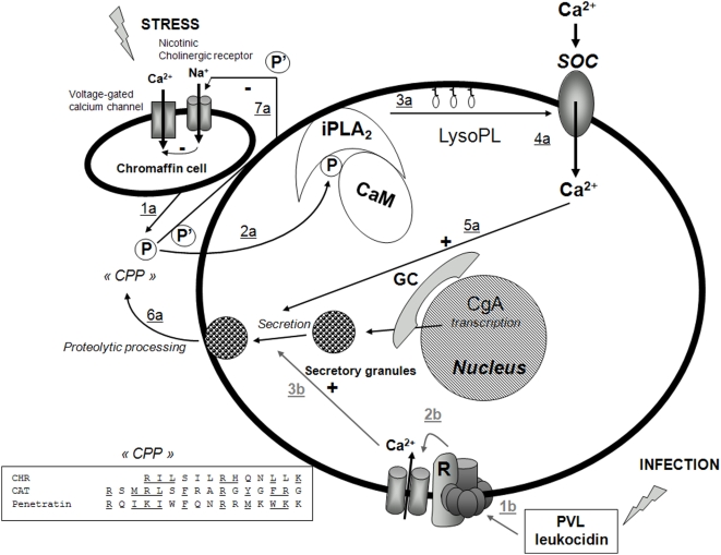
CONCLUSIONS/SIGNIFICANCE: For the first time, we demonstrate that CHR and CAT, penetrate into PMNs, inducing extracellular calcium entry by a CaM-regulated iPLA2 pathway. Our study highlights the role of two CgA-derived peptides in the active communication between neuroendocrine and immune systems.
嗜铬粒蛋白A(CgA)自然加工产生的抗菌肽在嗜铬细胞受刺激时与儿茶酚胺共同分泌。由于中性粒细胞在先天免疫中起核心作用,我们研究了两种抗菌CgA衍生肽刺激后中性粒细胞的反应。
方法/主要发现:在几种药物存在的情况下,用不同浓度的CgA衍生肽处理中性粒细胞。通过流式细胞术和钙成像实验观察钙动员情况。免疫细胞化学和共聚焦显微镜显示了肽的细胞内定位。通过表面等离子体共振和iPLA2活性测定显示了肽的钙调蛋白结合和iPLA2激活特性。最后,通过使用HPLC和纳米液相色谱-质谱联用仪对用CgA衍生肽处理后的中性粒细胞释放的物质进行蛋白质组分析。通过流式细胞术,我们首先观察到,在细胞外钙存在的情况下,15秒后,嗜铬菌素(CHR)或抑胃肽(CAT)会引起细胞内钙浓度依赖性的瞬时增加。相反,在没有细胞外钙的情况下,暴露10分钟后,这些肽无法诱导细胞内钙库的钙消耗。如钙成像所示,用2-氨基乙氧基二苯硼酸盐(2-APB)(一种储存操纵通道(SOCs)阻滞剂)处理可完全抑制钙内流。我们还表明,它们作为两个钙调蛋白结合因子(W7和CMZ)激活iPLA2,并且这两个序列可以与报道的iPLA2的两个钙调蛋白结合结构域对齐。我们最后通过HPLC和纳米液相色谱-质谱联用仪分析了CHR和CAT刺激后中性粒细胞释放的物质。我们鉴定了几种对炎症和先天免疫很重要的因子。
结论/意义:我们首次证明,CHR和CAT可穿透中性粒细胞,通过钙调蛋白调节的iPLA2途径诱导细胞外钙内流。我们的研究突出了两种CgA衍生肽在神经内分泌系统和免疫系统之间主动通讯中的作用。