• 文献检索
  • 文档翻译
  • 深度研究
  • 学术资讯
  • Suppr Zotero 插件Zotero 插件
  • 邀请有礼
  • 套餐&价格
  • 历史记录
应用&插件
Suppr Zotero 插件Zotero 插件浏览器插件Mac 客户端Windows 客户端微信小程序
定价
高级版会员购买积分包购买API积分包
服务
文献检索文档翻译深度研究API 文档MCP 服务
关于我们
关于 Suppr公司介绍联系我们用户协议隐私条款
关注我们

Suppr 超能文献

核心技术专利:CN118964589B侵权必究
粤ICP备2023148730 号-1Suppr @ 2026

文献检索

告别复杂PubMed语法,用中文像聊天一样搜索,搜遍4000万医学文献。AI智能推荐,让科研检索更轻松。

立即免费搜索

文件翻译

保留排版,准确专业,支持PDF/Word/PPT等文件格式,支持 12+语言互译。

免费翻译文档

深度研究

AI帮你快速写综述,25分钟生成高质量综述,智能提取关键信息,辅助科研写作。

立即免费体验

犬腕管的超声检查

Ultrasonographic examination of the carpal canal in dogs.

作者信息

Turan Erkut, Ozsunar Yelda, Yildirim Ismail Gokce

机构信息

Department of Anatomy, Faculty of Veterinary Medicine, University of Adnan Menderes, PK: 17, 09016, Isikli-Aydin, Turkey.

出版信息

J Vet Sci. 2009 Mar;10(1):77-80. doi: 10.4142/jvs.2009.10.1.77.

DOI:10.4142/jvs.2009.10.1.77
PMID:19255528
原文链接:https://pmc.ncbi.nlm.nih.gov/articles/PMC2801093/
Abstract

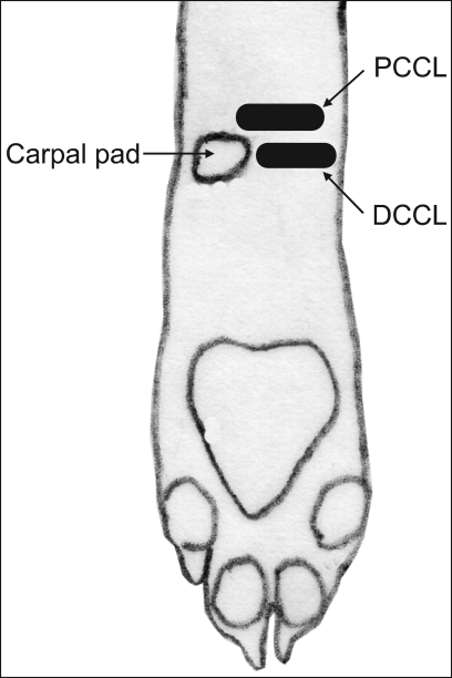
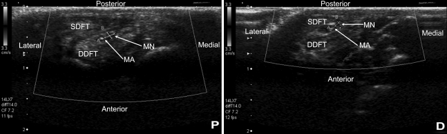
The aim of this study was to determine the course of the median nerve and its adjacent structures in the carpal canals of 8 healthy dogs by using high-frequency transducers. Before performing ultrasonography, the transverse and posteroanterior diameters as well as the perimeter of the carpus were measured at just proximal to the side of the carpal pad. The anatomical structures were then determined at two levels of the carpal canal, which were named the proximal and distal levels, on the transverse sonograms. The cross-sectional areas, perimeters and the transverse and posteroanterior diameters of the median nerve were measured at these levels. Although all the measurements were larger at the proximal level, significant differences between the proximal and distal levels were determined for the cross-sectional area, the perimeter and the transverse diameter of the median nerve. On the transverse sonogram, the deep digital flexor tendon was seen in almost the center of the carpal canal like a comma shape and also it had a small concavity on the caudal side. The superficial digital flexor tendon was seen as an ovoid shape on the transverse sonograms and it was located nearly at the posterior side of the carpal canal. Both tendons were seen as intermediate-grade echogenic structures. The median artery was located inside of the concavity of the deep digital flexor tendon. Also, the median nerve was seen at the posteromedial side of the median artery. As a result of this study, the cross-sectional areas of the median nerve ranged between 1.01-2.68 mm(2) at the proximal level and between 0.93-1.91 mm(2) at the distal level.

摘要

本研究的目的是通过使用高频换能器来确定8只健康犬腕管内正中神经及其相邻结构的走行。在进行超声检查之前,于腕垫一侧近端测量腕关节的横径、前后径以及周长。然后在横断超声图像上,于腕管的两个水平(分别命名为近端水平和远端水平)确定解剖结构。在这些水平测量正中神经的横截面积、周长以及横径和前后径。尽管所有测量值在近端水平均更大,但正中神经的横截面积、周长和横径在近端和远端水平之间存在显著差异。在横断超声图像上,指深屈肌腱在腕管几乎中央处呈逗号形可见,并且在尾侧有一小凹陷。指浅屈肌腱在横断超声图像上呈椭圆形可见,且位于腕管后侧附近。两条肌腱均呈中等回声结构。正中动脉位于指深屈肌腱凹陷内。此外,正中神经在正中动脉的后内侧可见。本研究结果显示,正中神经的横截面积在近端水平为1.01 - 2.68平方毫米,在远端水平为0.93 - 1.91平方毫米。

https://cdn.ncbi.nlm.nih.gov/pmc/blobs/01bf/2801093/a6e5bf93c270/jvs-10-77-g002.jpg
https://cdn.ncbi.nlm.nih.gov/pmc/blobs/01bf/2801093/16a385719949/jvs-10-77-g001.jpg
https://cdn.ncbi.nlm.nih.gov/pmc/blobs/01bf/2801093/a6e5bf93c270/jvs-10-77-g002.jpg
https://cdn.ncbi.nlm.nih.gov/pmc/blobs/01bf/2801093/16a385719949/jvs-10-77-g001.jpg
https://cdn.ncbi.nlm.nih.gov/pmc/blobs/01bf/2801093/a6e5bf93c270/jvs-10-77-g002.jpg

相似文献

1
Ultrasonographic examination of the carpal canal in dogs.犬腕管的超声检查
J Vet Sci. 2009 Mar;10(1):77-80. doi: 10.4142/jvs.2009.10.1.77.
2
Computed tomography and morphometry of the carpal canal in the dog.犬腕管的计算机断层扫描和形态测量
Ann Anat. 2003 Apr;185(2):173-8. doi: 10.1016/S0940-9602(03)80084-5.
3
Ultrasonographic cross-sectional area and mean echogenicity of the superficial and deep digital flexor tendons in 50 trained thoroughbred racehorses.50匹训练有素的纯种赛马的指浅屈肌腱和指深屈肌腱的超声横截面积及平均回声强度
Am J Vet Res. 1995 Oct;56(10):1265-9.
4
A Case Series of Five Horses with Superficial Digital Flexor Tendon Lesions in the Carpal Canal.腕管内浅层指屈肌腱病变的 5 例马病例系列
J Equine Vet Sci. 2021 Aug;103:103656. doi: 10.1016/j.jevs.2021.103656. Epub 2021 May 11.
5
Ultrasonography as a method to determine tendon cross-sectional area.超声检查作为一种确定肌腱横截面积的方法。
Am J Vet Res. 1995 Oct;56(10):1270-4.
6
Comparison of proximal and distal cross-sectional areas of the median nerve, carpal tunnel, and nerve/tunnel index in subjects with carpal tunnel syndrome.正中神经近端和远端横截面积、腕管以及腕管综合征患者正中神经/腕管指数的比较。
Arch Phys Med Rehabil. 2013 Nov;94(11):2151-6. doi: 10.1016/j.apmr.2013.05.008. Epub 2013 May 28.
7
Gross and ultrasonographic anatomy of the carpal flexor tendon sheath in horses.
Vet Rec. 1997 Nov 8;141(19):489-95. doi: 10.1136/vr.141.19.489.
8
Ultrasonographic anatomy of the palmar region of the carpus of the dog.犬腕掌侧超声解剖。
Vet Radiol Ultrasound. 2023 May;64(3):546-556. doi: 10.1111/vru.13224. Epub 2023 Mar 7.
9
Tenoscopic anatomy of the equine carpal flexor synovial sheath.马腕部屈肌滑膜鞘的腱鞘解剖
Vet Surg. 1998 Mar-Apr;27(2):150-7. doi: 10.1111/j.1532-950x.1998.tb00112.x.
10
Desmotomy of the accessory ligament of the superficial digital flexor tendon in the horse with use of a tenoscopic approach to the carpal sheath.采用腱鞘镜技术对马的指浅屈肌腱副韧带进行离断术。
Vet Surg. 1999 Mar-Apr;28(2):99-105. doi: 10.1053/jvet.1999.0099.

引用本文的文献

1
Anatomy of the Palmar Region of the Carpus of the Dog.犬腕掌侧区域的解剖结构
Animals (Basel). 2022 Jun 18;12(12):1573. doi: 10.3390/ani12121573.
2
The Structure of the Brachial Plexus in Selected Representatives of the Caniformia Suborder.犬形亚目部分代表动物的臂丛神经结构
Animals (Basel). 2022 Feb 23;12(5):566. doi: 10.3390/ani12050566.
3
Clinical Applications of Imaging Modalities of the Carpal Joint in Dogs with Particular Reference to the Carpal Canal.犬腕关节成像方式的临床应用,特别涉及腕管

本文引用的文献

1
Ultrasonographic assessment of the canine sciatic nerve.犬坐骨神经的超声评估
Vet Radiol Ultrasound. 2007 Sep-Oct;48(5):428-33. doi: 10.1111/j.1740-8261.2007.00273.x.
2
Ultrasonographic evaluation of brachial plexus tumors in five dogs.五只犬臂丛神经肿瘤的超声评估
Vet Radiol Ultrasound. 2005 Nov-Dec;46(6):514-7. doi: 10.1111/j.1740-8261.2005.00093.x.
3
Ultrasonographic anatomy of the canine elbow.犬肘部的超声解剖学
J Vet Res. 2020 Mar 11;64(1):169-174. doi: 10.2478/jvetres-2020-0006. eCollection 2020 Mar.
Vet Radiol Ultrasound. 2005 Jul-Aug;46(4):319-25. doi: 10.1111/j.1740-8261.2005.00060.x.
4
Diagnostic value of sonography in patients with suspected carpal tunnel syndrome: a prospective study.超声检查对疑似腕管综合征患者的诊断价值:一项前瞻性研究。
Arthritis Rheum. 2005 Jan;52(1):304-11. doi: 10.1002/art.20723.
5
Correlation of high-resolution ultrasonographic findings with the clinical symptoms and electrodiagnostic data in carpal tunnel syndrome.腕管综合征中高分辨率超声检查结果与临床症状及电诊断数据的相关性
Ann Plast Surg. 2005 Jan;54(1):20-3. doi: 10.1097/01.sap.0000141942.27182.55.
6
The role of ultrasonographic measurements of the median nerve in the diagnosis of carpal tunnel syndrome.正中神经超声测量在腕管综合征诊断中的作用。
Clin Radiol. 2004 Oct;59(10):910-5. doi: 10.1016/j.crad.2004.03.020.
7
Evaluation of possible carpal tunnel syndrome in dogs.犬腕管综合征可能性的评估
Vet Rec. 2004 Jul 24;155(4):122-4. doi: 10.1136/vr.155.4.122.
8
High-resolution sonography of the peripheral nervous system -- a review of the literature.外周神经系统的高分辨率超声检查——文献综述
Eur J Neurol. 2004 May;11(5):305-14. doi: 10.1111/j.1468-1331.2004.00773.x.
9
Ultrasonography versus nerve conduction study in patients with carpal tunnel syndrome: substantive or complementary tests?腕管综合征患者的超声检查与神经传导研究:实质性检查还是补充性检查?
Rheumatology (Oxford). 2004 Jul;43(7):887-95. doi: 10.1093/rheumatology/keh190. Epub 2004 Apr 20.
10
Ultrasound of nerve and muscle.神经与肌肉的超声检查
Clin Neurophysiol. 2004 Mar;115(3):495-507. doi: 10.1016/j.clinph.2003.10.022.