Zanetti Marcus V, Jackowski Marcel P, Versace Amelia, Almeida Jorge R C, Hassel Stefanie, Duran Fábio L S, Busatto Geraldo F, Kupfer David J, Phillips Mary L
Laboratory of Psychiatric Neuroimaging (LIM-21), Department and Institute of Psychiatry, University of São Paulo Medical School, Centro de Medicina Nuclear, São Paulo, SP 05403-010, Brazil.
Eur Arch Psychiatry Clin Neurosci. 2009 Sep;259(6):316-28. doi: 10.1007/s00406-009-0002-8. Epub 2009 Mar 3.
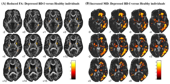
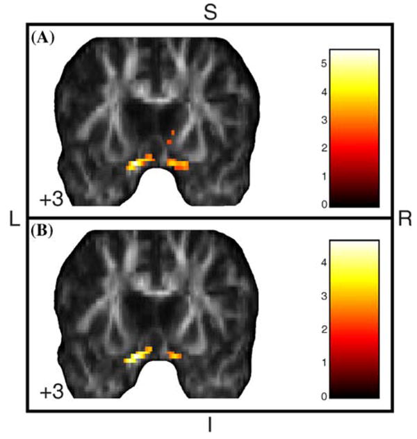
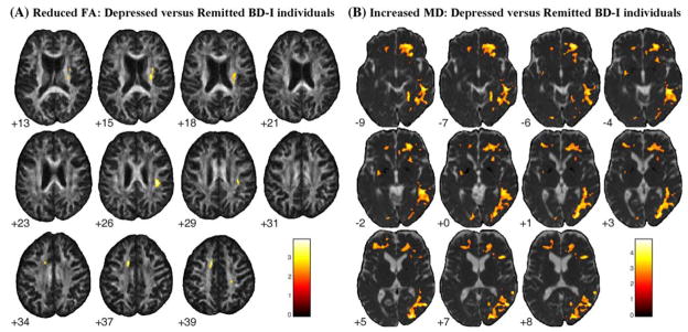
Abnormalities in fronto-limbic-striatal white matter (WM) have been reported in bipolar disorder (BD), but results have been inconsistent across studies. Furthermore, there have been no detailed investigations as to whether acute mood states contribute to microstructural changes in WM tracts. In order to compare fiber density and structural integrity within WM tracts between BD depression and remission, whole-brain fractional anisotropy (FA) and mean diffusivity (MD) were assessed in 37 bipolar I disorder (BD-I) patients (16 depressed and 21 remitted), and 26 healthy individuals with diffusion tensor imaging. Significantly decreased FA and increased MD in bilateral prefronto-limbic-striatal white matter and right inferior fronto-occipital, superior and inferior longitudinal fasciculi were shown in all BD-I patients versus controls, as well as in depressed BD-I patients compared to both controls and remitted BD-I patients. Depressed BD-I patients also exhibited increased FA in the ventromedial prefrontal cortex. Remitted BD-I patients did not differ from controls in FA or MD. These findings suggest that BD-I depression may be associated with acute microstructural WM changes.
双相情感障碍(BD)患者的额-边缘-纹状体白质(WM)异常已有报道,但各研究结果并不一致。此外,关于急性情绪状态是否会导致WM束的微观结构变化,尚未有详细研究。为比较BD抑郁发作期与缓解期WM束内的纤维密度和结构完整性,对37例双相I型障碍(BD-I)患者(16例抑郁发作期和21例缓解期)及26名健康个体进行了扩散张量成像,评估全脑分数各向异性(FA)和平均扩散率(MD)。与对照组相比,所有BD-I患者双侧额-边缘-纹状体白质以及右侧额枕下、上纵束和下纵束的FA显著降低,MD增加;与对照组和缓解期BD-I患者相比,抑郁发作期BD-I患者也有同样表现。抑郁发作期BD-I患者腹内侧前额叶皮质的FA也增加。缓解期BD-I患者的FA或MD与对照组无差异。这些发现表明,BD-I抑郁发作可能与急性WM微观结构变化有关。