Remme C A, Verkerk A O, Hoogaars W M H, Aanhaanen W T J, Scicluna B P, Annink C, van den Hoff M J B, Wilde A A M, van Veen T A B, Veldkamp M W, de Bakker J M T, Christoffels V M, Bezzina C R
Heart Failure Research Center, Academic Medical Center, University of Amsterdam, Amsterdam, The Netherlands.
Basic Res Cardiol. 2009 Sep;104(5):511-22. doi: 10.1007/s00395-009-0012-8. Epub 2009 Mar 3.
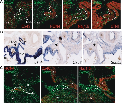
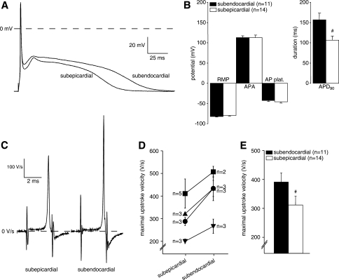
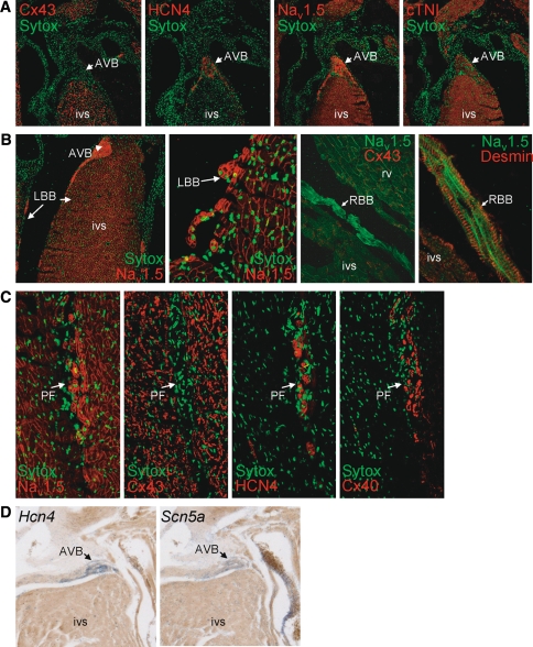
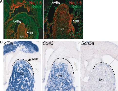
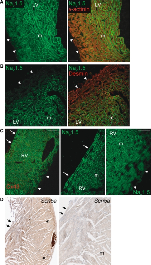
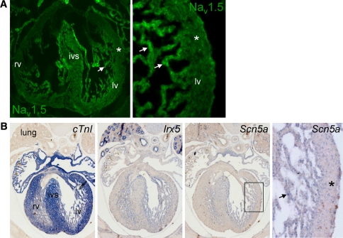
Cardiac sodium channels are responsible for conduction in the normal and diseased heart. We aimed to investigate regional and transmural distribution of sodium channel expression and function in the myocardium. Sodium channel Scn5a mRNA and Na(v)1.5 protein distribution was investigated in adult and embryonic mouse heart through immunohistochemistry and in situ hybridization. Functional sodium channel availability in subepicardial and subendocardial myocytes was assessed using patch-clamp technique. Adult and embryonic (ED14.5) mouse heart sections showed low expression of Na(v)1.5 in the HCN4-positive sinoatrial and atrioventricular nodes. In contrast, high expression levels of Na(v)1.5 were observed in the HCN4-positive and Cx43-negative AV or His bundle, bundle branches and Purkinje fibers. In both ventricles, a transmural gradient was observed, with a low Na(v)1.5 labeling intensity in the subepicardium as compared to the subendocardium. Similar Scn5a mRNA expression patterns were observed on in situ hybridization of embryonic and adult tissue. Maximal action potential upstroke velocity was significantly lower in subepicardial myocytes (mean +/- SEM 309 +/- 32 V/s; n = 14) compared to subendocardial myocytes (394 +/- 32 V/s; n = 11; P < 0.05), indicating decreased sodium channel availability in subepicardium compared to subendocardium. Scn5a and Na(v)1.5 show heterogeneous distribution patterns within the cardiac conduction system and across the ventricular wall. This differential distribution of the cardiac sodium channel may have profound consequences for conduction disease phenotypes and arrhythmogenesis in the setting of sodium channel disease.
心脏钠通道负责正常和患病心脏的电传导。我们旨在研究心肌中钠通道表达和功能的区域及透壁分布。通过免疫组织化学和原位杂交技术,研究了成年和胚胎小鼠心脏中钠通道Scn5a mRNA和Na(v)1.5蛋白的分布。使用膜片钳技术评估心外膜下和心内膜下心肌细胞中功能性钠通道的可用性。成年和胚胎(胚胎第14.5天)小鼠心脏切片显示,在HCN4阳性的窦房结和房室结中,Na(v)1.5表达较低。相反,在HCN4阳性且Cx43阴性的房室或希氏束、束支和浦肯野纤维中观察到Na(v)1.5的高表达水平。在两个心室中,均观察到透壁梯度,与心内膜相比,心外膜下Na(v)1.5标记强度较低。在胚胎和成年组织的原位杂交中观察到类似的Scn5a mRNA表达模式。与心内膜下心肌细胞(394 +/- 32 V/s;n = 11;P < 0.05)相比,心外膜下心肌细胞的最大动作电位上升速度显著更低(平均 +/- 标准误 309 +/- 32 V/s;n = 14),表明心外膜下钠通道可用性低于心内膜下。Scn5a和Na(v)1.5在心脏传导系统内和整个心室壁上显示出异质性分布模式。心脏钠通道的这种差异分布可能对钠通道疾病背景下的传导疾病表型和心律失常发生具有深远影响。