Wilks D C, Winwood K, Gilliver S F, Kwiet A, Chatfield M, Michaelis I, Sun L W, Ferretti J L, Sargeant A J, Felsenberg D, Rittweger J
Institute for Biomedical Research into Human Movement and Health, Manchester Metropolitan University, Manchester, UK.
Bone. 2009 Jul;45(1):91-7. doi: 10.1016/j.bone.2009.03.660. Epub 2009 Mar 28.
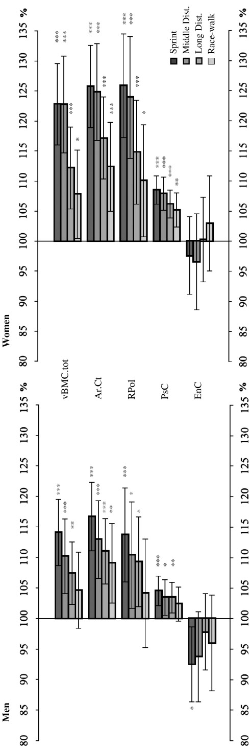
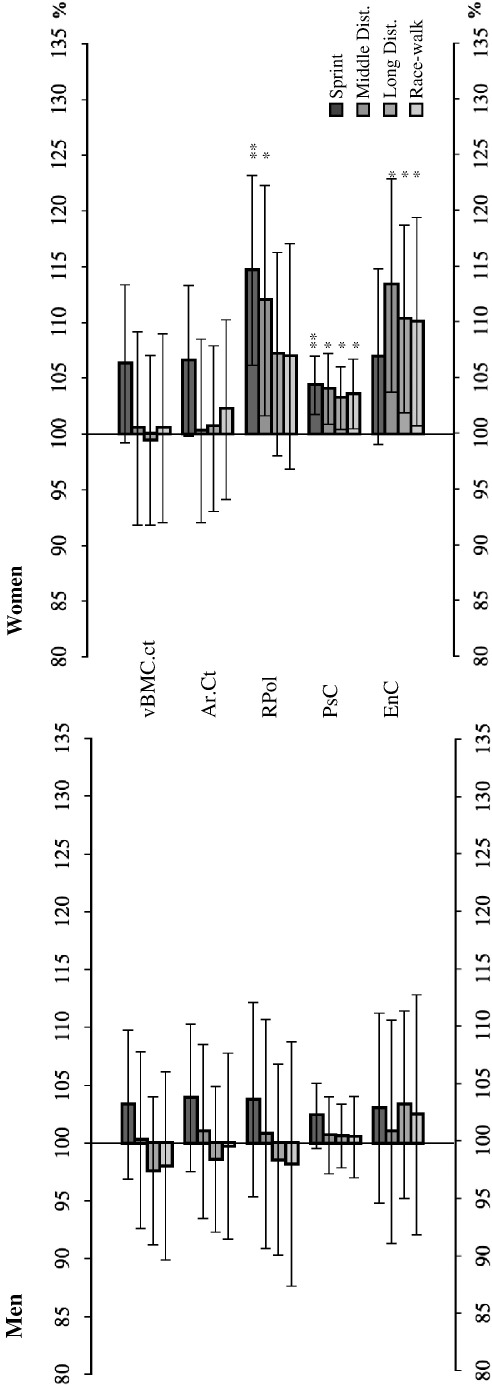
Mechanical loading is thought to be a determinant of bone mass and geometry. Both ground reaction forces and tibial strains increase with running speed. This study investigates the hypothesis that surrogates of bone strength in male and female master sprinters, middle and long distance runners and race-walkers vary according to discipline-specific mechanical loading from sedentary controls. Bone scans were obtained by peripheral Quantitative Computed Tomography (pQCT) from the tibia and from the radius in 106 sprinters, 52 middle distance runners, 93 long distance runners and 49 race-walkers who were competing at master championships, and who were aged between 35 and 94 years. Seventy-five age-matched, sedentary people served as control group. Most athletes of this study had started to practice their athletic discipline after the age of 20, but the current training regime had typically been maintained for more than a decade. As hypothesised, tibia diaphyseal bone mineral content (vBMC), cortical area and polar moment of resistance were largest in sprinters, followed in descending order by middle and long distance runners, race-walkers and controls. When compared to control people, the differences in these measures were always >13% in male and >23% in female sprinters (p<0.001). Similarly, the periosteal circumference in the tibia shaft was larger in male and female sprinters by 4% and 8%, respectively, compared to controls (p<0.001). Epiphyseal group differences were predominantly found for trabecular vBMC in both male and female sprinters, who had 15% and 18% larger values, respectively, than controls (p<0.001). In contrast, a reverse pattern was found for cortical vBMD in the tibia, and only few group differences of lower magnitude were found between athletes and control people for the radius. In conclusion, tibial bone strength indicators seemed to be related to exercise-specific peak forces, whilst cortical density was inversely related to running distance. These results may be explained in two, non-exclusive ways. Firstly, greater skeletal size may allow larger muscle forces and power to be exerted, and thus bias towards engagement in athletics. Secondly, musculoskeletal forces related to running can induce skeletal adaptation and thus enhance bone strength.
机械负荷被认为是骨量和骨几何结构的一个决定因素。地面反作用力和胫骨应变均随跑步速度的增加而增大。本研究调查了这样一个假设:男女成年短跑运动员、中长跑运动员和竞走运动员的骨强度替代指标会因与久坐对照组不同的特定项目机械负荷而有所差异。通过外周定量计算机断层扫描(pQCT)对106名短跑运动员、52名中长跑运动员、93名长跑运动员和49名竞走运动员的胫骨和桡骨进行骨扫描,这些运动员均参加成年组锦标赛,年龄在35至94岁之间。75名年龄匹配的久坐者作为对照组。本研究中的大多数运动员在20岁以后开始从事其运动项目,但目前的训练模式通常已维持了十多年。正如所假设的那样,胫骨骨干骨矿物质含量(vBMC)、皮质面积和抗扭极矩在短跑运动员中最大,其次依次是中长跑运动员、竞走运动员和对照组。与对照组相比,男性和女性短跑运动员在这些指标上的差异始终分别大于13%和23%(p<0.001)。同样,与对照组相比,男性和女性短跑运动员胫骨骨干的骨膜周长分别增大了4%和8%(p<0.001)。在男女短跑运动员的骨骺组中,主要发现小梁vBMC存在差异,其值分别比对照组大15%和18%(p<0.001)。相比之下,在胫骨皮质骨密度方面发现了相反的模式,并且在运动员和对照组之间,桡骨仅发现了少数较小幅度的组间差异。总之,胫骨骨强度指标似乎与特定运动的峰值力有关,而皮质骨密度与跑步距离呈负相关。这些结果可以用两种并非相互排斥的方式来解释。首先,更大的骨骼尺寸可能使更大的肌肉力量和功率得以发挥,从而偏向于从事体育运动。其次,与跑步相关的肌肉骨骼力可以诱导骨骼适应,从而增强骨强度。