Artang Ramin, Migrino Raymond Q, Harmann Leanne, Bowers Mark, Woods Timothy D
Division of Cardiovascular Medicine, Medical College of Wisconsin, Milwaukee, Wisconsin, USA.
Cardiovasc Ultrasound. 2009 Mar 31;7:16. doi: 10.1186/1476-7120-7-16.
Left atrial size is an important marker for adverse cardiovascular events. There is general consensus that left atrial volume index (LAVI) is the best measurement of size. The current LAVI measurement techniques are laborious. Semi-automated measurement with a 3-dimensional echocardiography (3DE) system may be a practical clinical alternative to measure LAVI, but it has not been adequately evaluated against Magnetic Resonance Imaging (MRI) gold standard. The aim of this study was to compare the accuracy of a commercially available 3D algorithm for measurement of LAVI against LAVI obtained from MRI and Area Length Method (ALM).
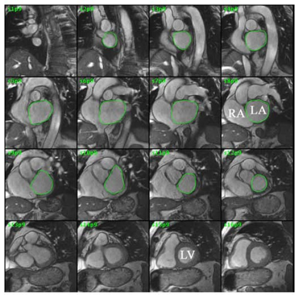
In 27 consecutive subjects referred for cardiac MRI (age 54 +/- 13 years, 63% male), LAVI was measured using 3 imaging modalities: 3DE, ALM, MRI and the results were correlated. ALM was measured using standard American Society of Echocardiography guidelines. The time required to measure LAVI by 3DE and ALM were compared.
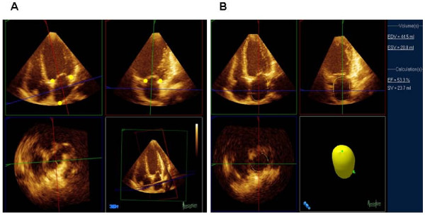
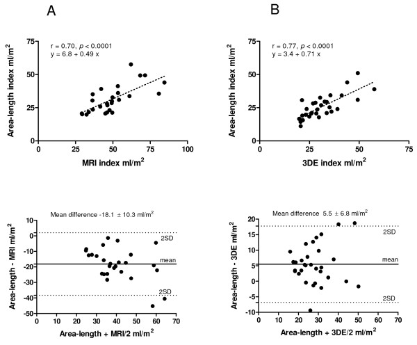
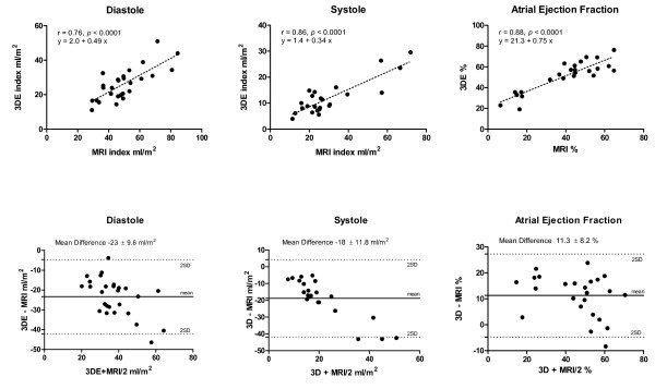
There was a significant correlation in systolic and diastolic LA volumes and left atrial ejection fraction between 3DE and MRI (r = 0.86 for systole, r = 0.76 for diastole, r = 0.88 for ejection fraction, P < 0.0001 for all). There was also significant correlation of diastolic volumes between 3DE and ALM (r = 0.77, P < 0.0001). The time to obtain LAVI was shorter using 3DE versus ALM (56 +/- 8 vs 135 +/- 55 seconds, P < 0.0001).
Three-dimensional echocardiography with semiautomatic border detection is a practical alternative for obtaining the left atrial volume in a time-efficient manner compared to the current standard.
左心房大小是心血管不良事件的重要标志物。人们普遍认为左心房容积指数(LAVI)是测量大小的最佳指标。当前的LAVI测量技术较为繁琐。使用三维超声心动图(3DE)系统进行半自动测量可能是临床上测量LAVI的一种实用替代方法,但与磁共振成像(MRI)金标准相比,其尚未得到充分评估。本研究的目的是比较一种商用3D算法测量LAVI的准确性与通过MRI和面积长度法(ALM)获得的LAVI的准确性。
在连续27例因心脏MRI检查而就诊的受试者中(年龄54±13岁,男性占63%),使用3种成像方式测量LAVI:3DE、ALM、MRI,并对结果进行相关性分析。使用美国超声心动图学会标准指南测量ALM。比较通过3DE和ALM测量LAVI所需的时间。
3DE与MRI之间在收缩期和舒张期左心房容积以及左心房射血分数方面存在显著相关性(收缩期r = 0.86,舒张期r = 0.76,射血分数r = 0.88,所有P < 0.0001)。3DE与ALM之间在舒张期容积方面也存在显著相关性(r = 0.77,P < 0.0001)。与ALM相比,使用3DE获得LAVI的时间更短(56±8秒对135±55秒,P < 0.0001)。
与当前标准相比,具有半自动边界检测功能的三维超声心动图是一种高效获取左心房容积的实用替代方法。