Walshe Tony E, Saint-Geniez Magali, Maharaj Arindel S R, Sekiyama Eiichi, Maldonado Angel E, D'Amore Patricia A
Department of Ophthalmology and Pathology, Harvard Medical School, Schepens Eye Research Institute, Boston, MA, USA.
PLoS One. 2009;4(4):e5149. doi: 10.1371/journal.pone.0005149. Epub 2009 Apr 2.
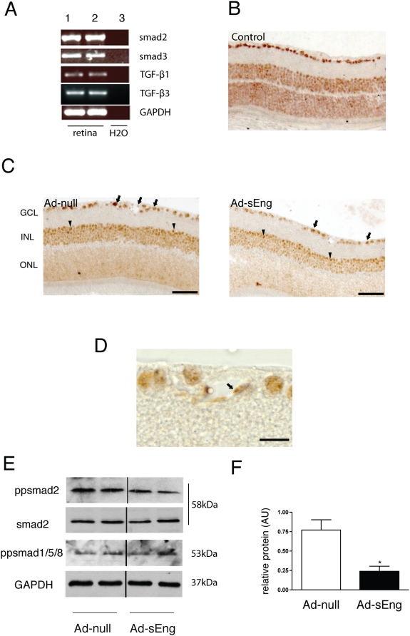
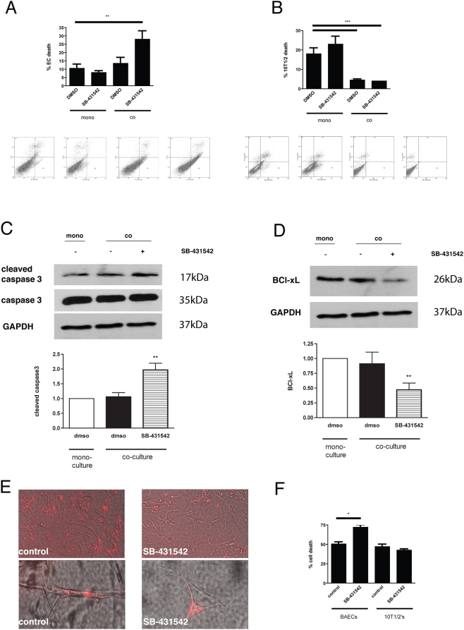
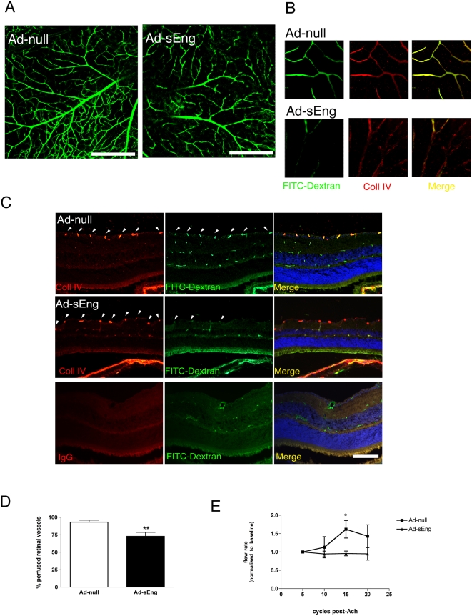
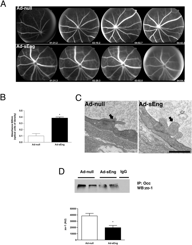
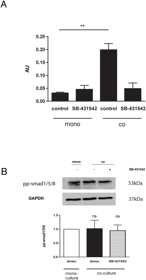
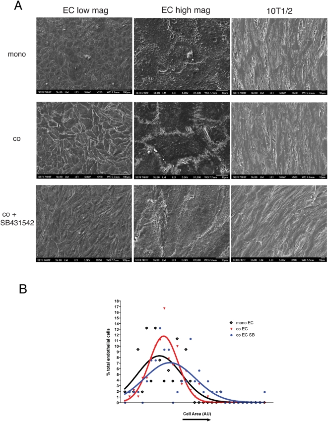
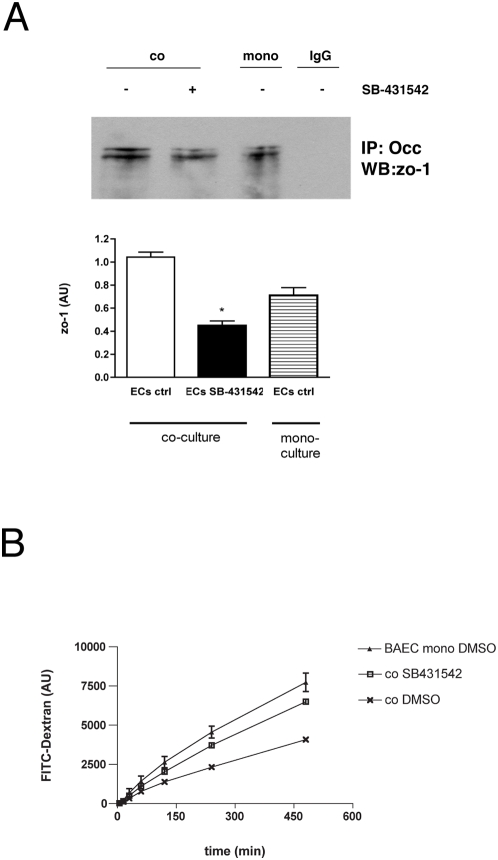
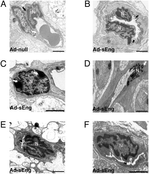
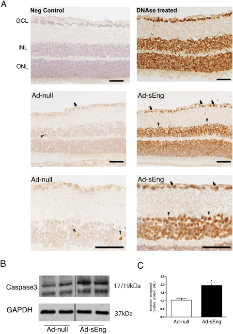
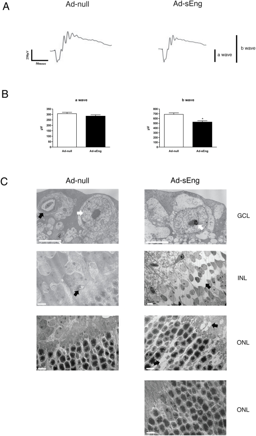
Pericyte-endothelial cell (EC) interactions are critical to both vascular development and vessel stability. We have previously shown that TGF-beta signaling between EC and mural cells participates in vessel stabilization in vitro. We therefore investigated the role of TGF-beta signaling in maintaining microvessel structure and function in the adult mouse retinal microvasculature. TGF-beta signaling was inhibited by systemic expression of soluble endoglin (sEng) and inhibition was demonstrated by reduced phospho-smad2 in the adult retina. Blockade of TGF-beta signaling led to increased vascular and neural cell apoptosis in the retina, which was associated with decreased retinal function, as measured by electroretinogram (ERG). Perfusion of the inner retinal vasculature was impaired and was accompanied by defective autoregulation and loss of capillary integrity. Fundus angiography and Evans blue permeability assay revealed a breakdown of the blood-retinal-barrier that was characterized by decreased association between the tight junction proteins zo-1 and occludin. Inhibition of TGF-beta signaling in cocultures of EC and 10T1/2 cells corroborated the in vivo findings, with impaired EC barrier function, dissociation of EC from 10T1/2 cells, and endothelial cell death, supporting the role of EC-mesenchymal interactions in TGF-beta signaling. These results implicate constitutive TGF-beta signaling in maintaining the integrity and function of the adult microvasculature and shed light on the potential role of TGF-beta signaling in vasoproliferative and vascular degenerative retinal diseases.
周细胞与内皮细胞(EC)的相互作用对于血管发育和血管稳定性都至关重要。我们之前已经表明,EC与壁细胞之间的TGF-β信号传导参与体外血管稳定。因此,我们研究了TGF-β信号传导在维持成年小鼠视网膜微血管系统微血管结构和功能中的作用。通过可溶性内皮糖蛋白(sEng)的全身表达抑制TGF-β信号传导,并且通过成年视网膜中磷酸化smad2的减少来证明抑制作用。阻断TGF-β信号传导导致视网膜中血管和神经细胞凋亡增加,这与视网膜电图(ERG)测量的视网膜功能下降有关。视网膜内血管系统的灌注受损,并伴有自调节缺陷和毛细血管完整性丧失。眼底血管造影和伊文思蓝通透性试验显示血视网膜屏障破坏,其特征是紧密连接蛋白zo-1和闭合蛋白之间的关联减少。在EC与10T1/2细胞共培养物中抑制TGF-β信号传导证实了体内研究结果,即EC屏障功能受损、EC与10T1/2细胞解离以及内皮细胞死亡,支持了EC-间充质相互作用在TGF-β信号传导中的作用。这些结果表明,组成型TGF-β信号传导在维持成年微血管系统的完整性和功能中起作用,并揭示了TGF-β信号传导在增殖性视网膜病变和血管退行性视网膜疾病中的潜在作用。