• 文献检索
  • 文档翻译
  • 深度研究
  • 学术资讯
  • Suppr Zotero 插件Zotero 插件
  • 邀请有礼
  • 套餐&价格
  • 历史记录
应用&插件
Suppr Zotero 插件Zotero 插件浏览器插件Mac 客户端Windows 客户端微信小程序
定价
高级版会员购买积分包购买API积分包
服务
文献检索文档翻译深度研究API 文档MCP 服务
关于我们
关于 Suppr公司介绍联系我们用户协议隐私条款
关注我们

Suppr 超能文献

核心技术专利:CN118964589B侵权必究
粤ICP备2023148730 号-1Suppr @ 2026

文献检索

告别复杂PubMed语法,用中文像聊天一样搜索,搜遍4000万医学文献。AI智能推荐,让科研检索更轻松。

立即免费搜索

文件翻译

保留排版,准确专业,支持PDF/Word/PPT等文件格式,支持 12+语言互译。

免费翻译文档

深度研究

AI帮你快速写综述,25分钟生成高质量综述,智能提取关键信息,辅助科研写作。

立即免费体验

用于三维体积重建的轨迹融合

Trajectory Fusion for Three-dimensional Volume Reconstruction.

作者信息

Lee Sang-Chul, Bajcsy Peter

机构信息

National Center for Supercomputing Applications, University of Illinois at Urbana-Champaign, 1205 W. Clark St, Urbana, IL 61801.

出版信息

Comput Vis Image Underst. 2008 Apr;110(1):19-31. doi: 10.1016/j.cviu.2007.02.005.

DOI:10.1016/j.cviu.2007.02.005
PMID:19343076
原文链接:https://pmc.ncbi.nlm.nih.gov/articles/PMC2387280/
Abstract

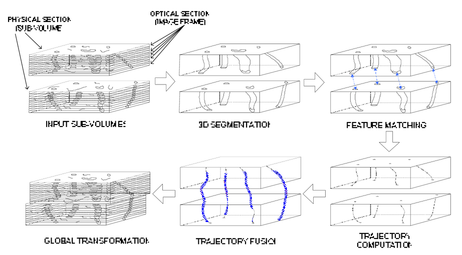
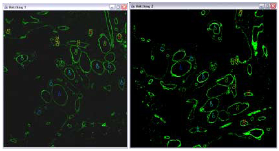
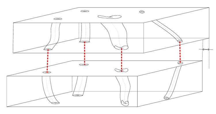
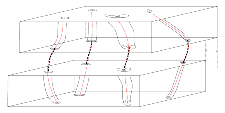
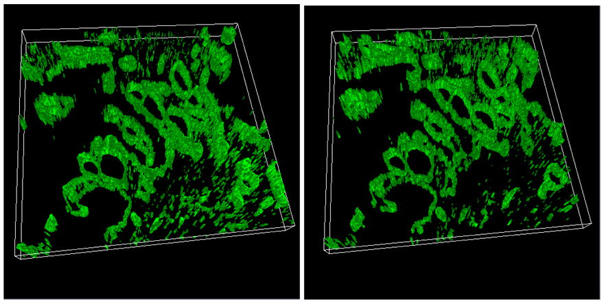
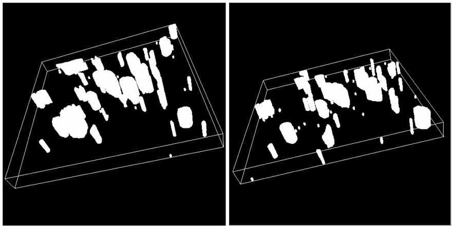
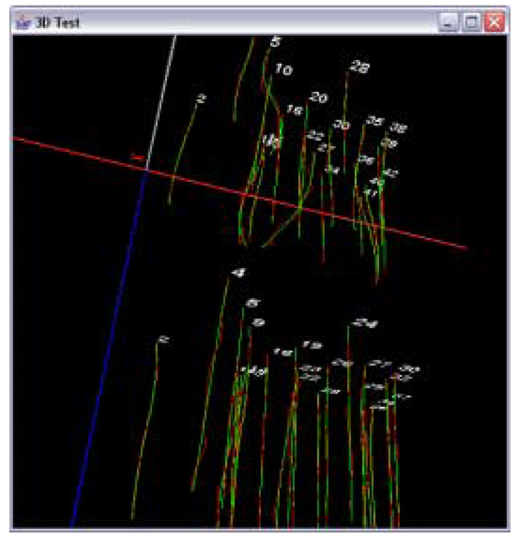
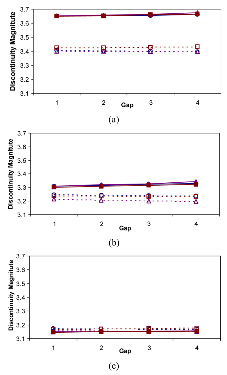
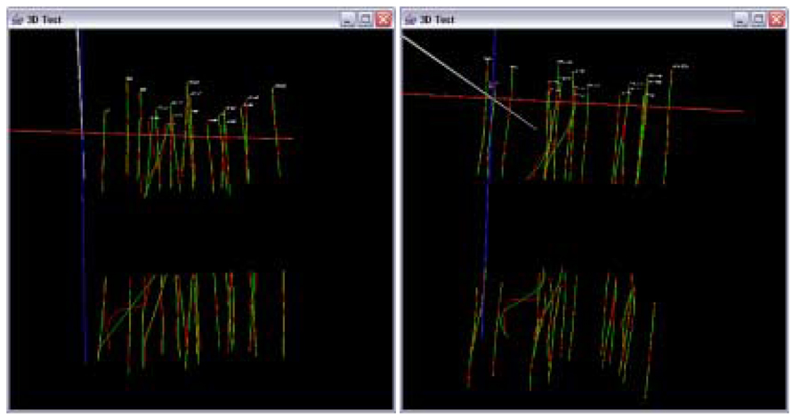
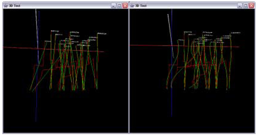
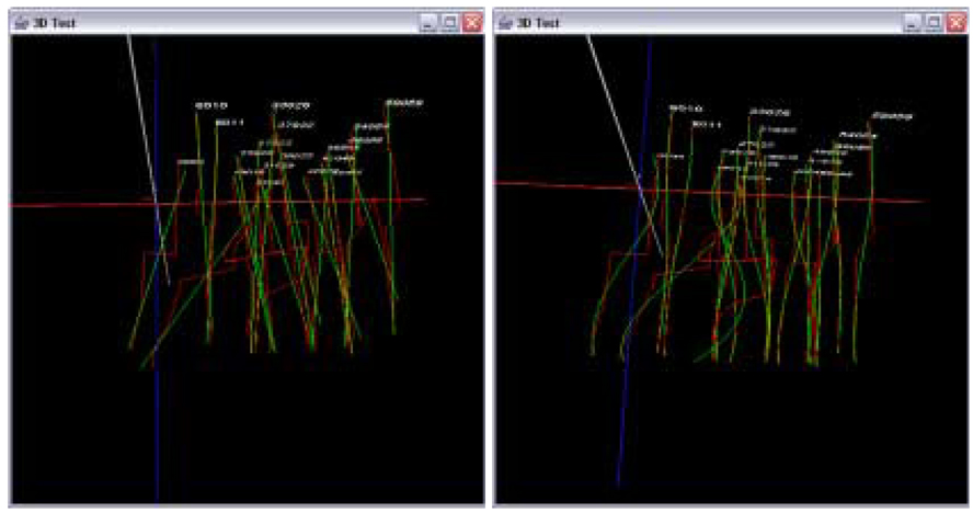
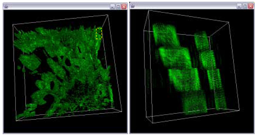
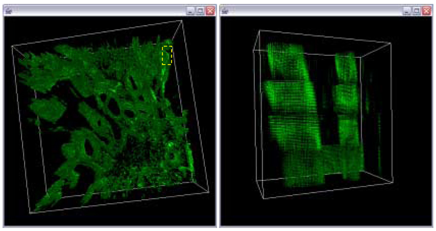
We address the 3D volume reconstruction problem from depth adjacent sub-volumes acquired by a confocal laser scanning microscope (CLSM). Our goal is to align the sub-volumes by estimating a set of optimal global transformations that preserve morphological continuity of medical structures, e.g., blood vessels, in the reconstructed 3D volume. We approach the problem by learning morphological characteristics of structures of interest in each sub-volume to understand global alignment transformations. Based on the observations of morphology, sub-volumes are aligned by connecting the morphological features at the sub-volume boundaries by minimizing morphological discontinuity. To minimize the discontinuity, we introduce three morphological discontinuity metrics: discontinuity magnitude at sub-volume boundary points, and overall and junction discontinuity residuals after polynomial curve fitting to multiple aligned sub-volumes. The proposed techniques have been applied to the problem of aligning CLSM sub-volumes acquired from four consecutive physical cross sections. Our experimental results demonstrated significant improvements of morphological smoothness of medical structures in comparison with the results obtained by feature matching at the sub-volume boundaries. The experimental results were evaluated by visual inspection and by quantifying morphological discontinuity metrics.

摘要

我们解决了由共聚焦激光扫描显微镜(CLSM)获取的深度相邻子体积的三维体积重建问题。我们的目标是通过估计一组最优的全局变换来对齐子体积,这些变换能保持重建三维体积中诸如血管等医学结构的形态连续性。我们通过学习每个子体积中感兴趣结构的形态特征来理解全局对齐变换,从而解决这个问题。基于形态学观察,通过最小化形态不连续性,在子体积边界处连接形态特征来对齐子体积。为了最小化不连续性,我们引入了三个形态不连续性度量:子体积边界点处的不连续幅度,以及对多个对齐子体积进行多项式曲线拟合后的整体和连接不连续残差。所提出的技术已应用于对齐从四个连续物理横截面获取的CLSM子体积的问题。与通过子体积边界处的特征匹配获得的结果相比,我们的实验结果表明医学结构的形态平滑度有显著提高。通过目视检查和量化形态不连续性度量对实验结果进行了评估。

https://cdn.ncbi.nlm.nih.gov/pmc/blobs/3165/2387280/b9534208c404/nihms44167f18.jpg
https://cdn.ncbi.nlm.nih.gov/pmc/blobs/3165/2387280/5a10c2a212d3/nihms44167f1.jpg
https://cdn.ncbi.nlm.nih.gov/pmc/blobs/3165/2387280/50a8c7a0f1d4/nihms44167f2.jpg
https://cdn.ncbi.nlm.nih.gov/pmc/blobs/3165/2387280/9a818930c2f0/nihms44167f3.jpg
https://cdn.ncbi.nlm.nih.gov/pmc/blobs/3165/2387280/b72ac9ce1639/nihms44167f4.jpg
https://cdn.ncbi.nlm.nih.gov/pmc/blobs/3165/2387280/1c3e9a45ed6f/nihms44167f5.jpg
https://cdn.ncbi.nlm.nih.gov/pmc/blobs/3165/2387280/fdc08ae3ab96/nihms44167f6.jpg
https://cdn.ncbi.nlm.nih.gov/pmc/blobs/3165/2387280/22c3896839f5/nihms44167f7.jpg
https://cdn.ncbi.nlm.nih.gov/pmc/blobs/3165/2387280/0645013ca7f1/nihms44167f8.jpg
https://cdn.ncbi.nlm.nih.gov/pmc/blobs/3165/2387280/e6054bf69962/nihms44167f9.jpg
https://cdn.ncbi.nlm.nih.gov/pmc/blobs/3165/2387280/0b3d312b4802/nihms44167f10a.jpg
https://cdn.ncbi.nlm.nih.gov/pmc/blobs/3165/2387280/2574dcc521cd/nihms44167f11.jpg
https://cdn.ncbi.nlm.nih.gov/pmc/blobs/3165/2387280/a169cf8e2a95/nihms44167f12a.jpg
https://cdn.ncbi.nlm.nih.gov/pmc/blobs/3165/2387280/8cd7d5b5c2cd/nihms44167f13.jpg
https://cdn.ncbi.nlm.nih.gov/pmc/blobs/3165/2387280/f1bdab9bcea3/nihms44167f14.jpg
https://cdn.ncbi.nlm.nih.gov/pmc/blobs/3165/2387280/7cf9793323d8/nihms44167f15.jpg
https://cdn.ncbi.nlm.nih.gov/pmc/blobs/3165/2387280/fc2d831fc842/nihms44167f16.jpg
https://cdn.ncbi.nlm.nih.gov/pmc/blobs/3165/2387280/ea462d978500/nihms44167f17.jpg
https://cdn.ncbi.nlm.nih.gov/pmc/blobs/3165/2387280/b9534208c404/nihms44167f18.jpg

相似文献

1
Trajectory Fusion for Three-dimensional Volume Reconstruction.用于三维体积重建的轨迹融合
Comput Vis Image Underst. 2008 Apr;110(1):19-31. doi: 10.1016/j.cviu.2007.02.005.
2
An intelligent workflow for sub-nanoscale 3D reconstruction of intact synapses from serial section electron tomography.用于从连续切片电子断层扫描对完整突触进行亚纳米级 3D 重建的智能工作流程。
BMC Biol. 2023 Sep 25;21(1):198. doi: 10.1186/s12915-023-01696-x.
3
Three-dimensional volume reconstruction of extracellular matrix proteins in uveal melanoma from fluorescent confocal laser scanning microscope images.利用荧光共聚焦激光扫描显微镜图像对葡萄膜黑色素瘤中细胞外基质蛋白进行三维体积重建。
J Microsc. 2006 Jan;221(Pt 1):30-45. doi: 10.1111/j.1365-2818.2006.01539.x.
4
Semi-automatic stitching of filamentous structures in image stacks from serial-section electron tomography.序列切片电子断层扫描图像堆栈中丝状结构的半自动拼接。
J Microsc. 2021 Oct;284(1):25-44. doi: 10.1111/jmi.13039. Epub 2021 Jul 9.
5
Automated multivolume placental reconstruction using three-dimensional power Doppler ultrasound and infrared camera tracking.使用三维能量多普勒超声和红外摄像机追踪技术进行自动多容积胎盘重建。
Ultrasound Obstet Gynecol. 2025 May;65(5):624-632. doi: 10.1002/uog.27708. Epub 2025 Apr 3.
6
Application of phase correlation to the montage synthesis and three-dimensional reconstruction of large tissue volumes from confocal laser scanning microscopy.相位相关在共聚焦激光扫描显微镜大组织体积的蒙太奇合成和三维重建中的应用。
Microsc Microanal. 2006 Apr;12(2):106-12. doi: 10.1017/S143192760606017X.
7
Enhancing panoramic dental imaging with AI-driven arch surface fitting: achieving improved clarity and accuracy through an optimal reconstruction zone.通过人工智能驱动的牙弓表面拟合增强全景牙科成像:通过优化重建区域实现更高的清晰度和准确性。
Dentomaxillofac Radiol. 2025 May 1;54(4):256-267. doi: 10.1093/dmfr/twaf006.
8
Semiautomatic segmentation of atherosclerotic carotid artery wall volume using 3D ultrasound imaging.使用三维超声成像对动脉粥样硬化颈动脉壁体积进行半自动分割。
Med Phys. 2015 Apr;42(4):2029-43. doi: 10.1118/1.4915925.
9
Accuracy Evaluation for Region Centroid-Based Registration of Fluorescent CLSM Imagery.基于区域质心的荧光共聚焦激光扫描显微镜图像配准的准确性评估
EURASIP J Appl Signal Processing. 2006;2006:1-11. doi: 10.1155/ASP/2006/82480.
10
Quantitative 3D Reconstruction from Scanning Electron Microscope Images Based on Affine Camera Models.基于仿射相机模型的扫描电子显微镜图像的定量 3D 重建。
Sensors (Basel). 2020 Jun 26;20(12):3598. doi: 10.3390/s20123598.

引用本文的文献

1
NeuroConstruct: 3D Reconstruction and Visualization of Neurites in Optical Microscopy Brain Images.NeuroConstruct:光学显微镜脑图像中神经突的 3D 重建与可视化。
IEEE Trans Vis Comput Graph. 2022 Dec;28(12):4951-4965. doi: 10.1109/TVCG.2021.3109460. Epub 2022 Oct 26.
2
Three-dimensional reconstruction of serial mouse brain sections: solution for flattening high-resolution large-scale mosaics.三维重建连续的小鼠脑切片:用于平铺高分辨率大尺度镶嵌图的解决方案。
Front Neuroanat. 2011 Mar 7;5:17. doi: 10.3389/fnana.2011.00017. eCollection 2011.

本文引用的文献

1
The local structure of image discontinuities in one dimension.一维图像不连续的局部结构。
IEEE Trans Pattern Anal Mach Intell. 1987 Mar;9(3):341-55. doi: 10.1109/tpami.1987.4767918.
2
Three-dimensional volume reconstruction of extracellular matrix proteins in uveal melanoma from fluorescent confocal laser scanning microscope images.利用荧光共聚焦激光扫描显微镜图像对葡萄膜黑色素瘤中细胞外基质蛋白进行三维体积重建。
J Microsc. 2006 Jan;221(Pt 1):30-45. doi: 10.1111/j.1365-2818.2006.01539.x.
3
First order error propagation of the procrustes method for 3D attitude estimation.
用于三维姿态估计的普罗克汝斯方法的一阶误差传播
IEEE Trans Pattern Anal Mach Intell. 2005 Feb;27(2):221-9. doi: 10.1109/TPAMI.2005.29.
4
Digital imaging fluorescence microscopy: spatial heterogeneity of photobleaching rate constants in individual cells.数字成像荧光显微镜:单个细胞中光漂白速率常数的空间异质性
J Cell Biol. 1985 Apr;100(4):1309-23. doi: 10.1083/jcb.100.4.1309.