• 文献检索
  • 文档翻译
  • 深度研究
  • 学术资讯
  • Suppr Zotero 插件Zotero 插件
  • 邀请有礼
  • 套餐&价格
  • 历史记录
应用&插件
Suppr Zotero 插件Zotero 插件浏览器插件Mac 客户端Windows 客户端微信小程序
定价
高级版会员购买积分包购买API积分包
服务
文献检索文档翻译深度研究API 文档MCP 服务
关于我们
关于 Suppr公司介绍联系我们用户协议隐私条款
关注我们

Suppr 超能文献

核心技术专利:CN118964589B侵权必究
粤ICP备2023148730 号-1Suppr @ 2026

文献检索

告别复杂PubMed语法,用中文像聊天一样搜索,搜遍4000万医学文献。AI智能推荐,让科研检索更轻松。

立即免费搜索

文件翻译

保留排版,准确专业,支持PDF/Word/PPT等文件格式,支持 12+语言互译。

免费翻译文档

深度研究

AI帮你快速写综述,25分钟生成高质量综述,智能提取关键信息,辅助科研写作。

立即免费体验

运动员左、右心室纵向峰值收缩期应变(斑点追踪测量)的实时评估。

Real-time evaluation of longitudinal peak systolic strain (speckle tracking measurement) in left and right ventricles of athletes.

作者信息

Stefani Laura, Pedrizzetti Gianni, De Luca Alessio, Mercuri Roberto, Innocenti Gabriele, Galanti Giorgio

机构信息

Sport Medicine Center, University of Florence, Italy.

出版信息

Cardiovasc Ultrasound. 2009 Apr 8;7:17. doi: 10.1186/1476-7120-7-17.

DOI:10.1186/1476-7120-7-17
PMID:19356233
原文链接:https://pmc.ncbi.nlm.nih.gov/articles/PMC2676253/
Abstract

BACKGROUND

Strain, and particularly Longitudinal Peak Systolic Strain (LPSS), plays a role in investigating the segmental and overall contractility of the heart which is a particularly interesting feature in athletes in whom regular training determines several morphological and functional modifications in both the ventricles, that normally work at different loads. Speckle tracking techniques assess the LPSS of LV and RV from B-mode imaging in real time, with uniform accuracy in all segments, and can verify the possible dissimilar segmental contributions of the two chambers to overall myocardial contraction. The aim of the study is to quantify the LPSS in real time in both the ventricles in order to estimate any possible different deformation properties in them during a systolic period.

METHODS

32 subjects (20 athletes and 18 controls) were submitted to a standard echocardiographic examination at rest and after a Hand Grip (HG) stress. From a four-chamber-view image, the LPSS parameter was measured with Speckle Tracking analysis in the basal and medium-apical segments of the two ventricles, at rest and after HG.

RESULTS

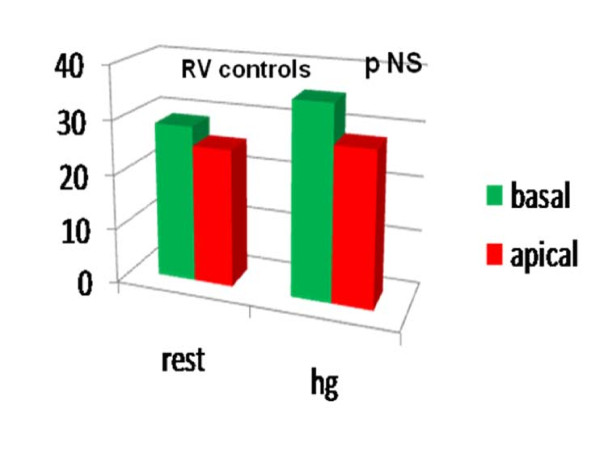
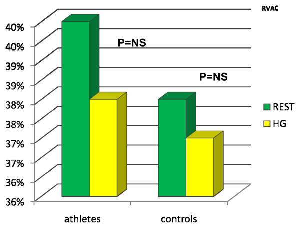
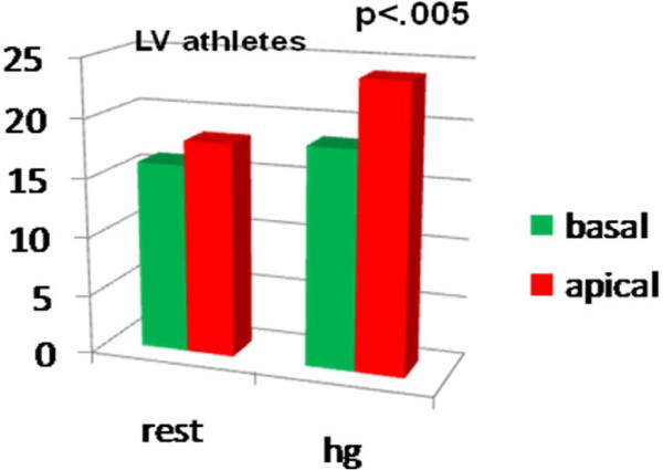
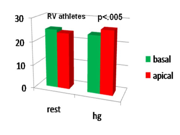
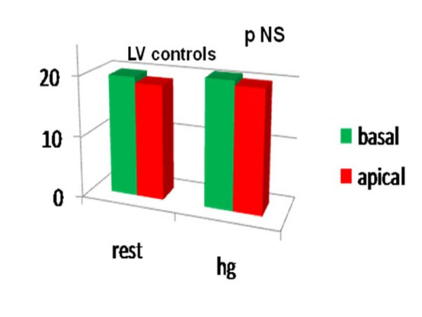
In both athletes and controls, LPSS values were significantly higher in the RV of athletes (RV LPSS medium-apical -23.87 +/- 4.94; basalfreewall -25.04 +/- 4.12 at rest) and controls (RV LPSSmedium-apical -25.21 +/- 4.97; basalfreewall -28.69 +/- 4.62 at rest) than in the LV of both (athletes LV LPSS medium-apical -18.14 +/- 4.16; basallateralwall -16.05 +/- 12.32; controls medium-apical -18.81 +/- 2.64; basallateralwall -19.74 +/- 3.84) With the HG test a significant enhancement of the LPSS(with P < .05) in the medium-apical segments of LV and RV was evident, but only in athletes; there was no modification of the standard echo-parameters in either group.

CONCLUSION

ST analysis is an easy method for investigating the contractility of the RV through deformation parameters, showing greater involvement of the RV than LV at rest. In athletes only, after isometric stress the two ventricles show particular myocardial deformation properties of the regions around the apex where the curvature of the wall is more marked. The clinical application of this new approach in athletes and normal subjects requires further investigation.

摘要

背景

应变,尤其是纵向峰值收缩应变(LPSS),在研究心脏节段性和整体收缩功能中发挥作用,这在运动员中是一个特别有趣的特征,因为规律训练会使两个心室发生多种形态和功能改变,而两个心室通常在不同负荷下工作。斑点追踪技术可从B模式成像实时评估左心室和右心室的LPSS,各节段准确性一致,且能验证两个心室对整体心肌收缩可能存在的不同节段贡献。本研究的目的是实时量化两个心室的LPSS,以估计收缩期它们中任何可能存在的不同变形特性。

方法

32名受试者(20名运动员和18名对照组)在静息状态和握力(HG)应激后接受标准超声心动图检查。从四腔心视图图像中,在静息状态和HG后,用斑点追踪分析测量两个心室基底段和中间心尖段的LPSS参数。

结果

在运动员和对照组中,运动员右心室的LPSS值(静息时右心室中间心尖段LPSS -23.87±4.94;基底游离壁 -25.04±4.12)和对照组(静息时右心室中间心尖段LPSS -25.21±4.97;基底游离壁 -28.69±4.62)均显著高于两者的左心室(运动员左心室中间心尖段LPSS -18.14±4.16;基底侧壁 -16.05±12.32;对照组中间心尖段 -18.81±2.64;基底侧壁 -19.74±3.84)。通过HG试验,左心室和右心室中间心尖段的LPSS有显著增强(P <.05),但仅在运动员中;两组的标准超声心动图均无改变。

结论

斑点追踪分析是一种通过变形参数研究右心室收缩功能的简便方法,显示静息时右心室比左心室受累更明显。仅在运动员中,等长应激后两个心室的心尖周围区域表现出特殊的心肌变形特性,此处壁的曲率更明显。这种新方法在运动员和正常受试者中的临床应用需要进一步研究。

https://cdn.ncbi.nlm.nih.gov/pmc/blobs/3008/2676253/ace712a011e0/1476-7120-7-17-7.jpg
https://cdn.ncbi.nlm.nih.gov/pmc/blobs/3008/2676253/47d9d6658f2e/1476-7120-7-17-1.jpg
https://cdn.ncbi.nlm.nih.gov/pmc/blobs/3008/2676253/ea63313fc7fe/1476-7120-7-17-2.jpg
https://cdn.ncbi.nlm.nih.gov/pmc/blobs/3008/2676253/c647e734931d/1476-7120-7-17-3.jpg
https://cdn.ncbi.nlm.nih.gov/pmc/blobs/3008/2676253/64ca363b9d5b/1476-7120-7-17-4.jpg
https://cdn.ncbi.nlm.nih.gov/pmc/blobs/3008/2676253/15c18c55f12b/1476-7120-7-17-5.jpg
https://cdn.ncbi.nlm.nih.gov/pmc/blobs/3008/2676253/e978a62420e9/1476-7120-7-17-6.jpg
https://cdn.ncbi.nlm.nih.gov/pmc/blobs/3008/2676253/ace712a011e0/1476-7120-7-17-7.jpg

相似文献

1
Real-time evaluation of longitudinal peak systolic strain (speckle tracking measurement) in left and right ventricles of athletes.运动员左、右心室纵向峰值收缩期应变(斑点追踪测量)的实时评估。
Cardiovasc Ultrasound. 2009 Apr 8;7:17. doi: 10.1186/1476-7120-7-17.
2
Comprehensive assessment of biventricular function and aortic stiffness in athletes with different forms of training by three-dimensional echocardiography and strain imaging.应用三维超声心动图和应变成像技术对不同形式训练的运动员的双心室功能和主动脉僵硬度进行综合评估。
Eur Heart J Cardiovasc Imaging. 2013 Oct;14(10):1010-20. doi: 10.1093/ehjci/jes298. Epub 2013 Jan 8.
3
Could quantitative longitudinal peak systolic strain help in the detection of left ventricular wall motion abnormalities in our daily echocardiographic practice?在我们日常的超声心动图检查实践中,定量纵向峰值收缩期应变能否有助于检测左心室壁运动异常?
Arch Cardiovasc Dis. 2016 Oct;109(10):533-541. doi: 10.1016/j.acvd.2016.02.007. Epub 2016 Jun 22.
4
Assessment of left ventricular systolic and diastolic abnormalities in patients with hypertrophic cardiomyopathy using real-time three-dimensional echocardiography and two-dimensional speckle tracking imaging.使用实时三维超声心动图和二维斑点追踪成像评估肥厚型心肌病患者的左心室收缩和舒张异常。
Cardiovasc Ultrasound. 2018 Oct 2;16(1):23. doi: 10.1186/s12947-018-0142-y.
5
Two-dimensional tracking and TDI are consistent methods for evaluating myocardial longitudinal peak strain in left and right ventricle basal segments in athletes.二维追踪和组织多普勒成像(TDI)是评估运动员左、右心室基底段心肌纵向峰值应变的一致方法。
Cardiovasc Ultrasound. 2007 Feb 7;5:7. doi: 10.1186/1476-7120-5-7.
6
Reproducibility of Combined Acquisition and Measurement of Left Ventricular Longitudinal Peak Segmental Strain in Relation to the Severity of Left Ventricular Dysfunction.左心室纵向节段峰值应变的联合获取和测量与左心室功能障碍严重程度的相关性的可重复性。
J Am Soc Echocardiogr. 2019 Nov;32(11):1451-1461.e3. doi: 10.1016/j.echo.2019.07.007. Epub 2019 Sep 23.
7
Right Ventricular Function and Region-Specific Adaptation in Athletes Engaged in High-Dynamic Sports: A Meta-Analysis.运动员从事高动态运动时的右心室功能和区域性适应:荟萃分析。
Circ Cardiovasc Imaging. 2021 May;14(5):e012315. doi: 10.1161/CIRCIMAGING.120.012315. Epub 2021 May 17.
8
Left and right ventricular morphology and function in athletes with elevated pulmonary systolic arterial pressure.肺动脉收缩压升高的运动员的左右心室形态与功能
Echocardiography. 2018 Jun;35(6):769-776. doi: 10.1111/echo.14016. Epub 2018 May 11.
9
Right ventricular remodelling induced by exercise training in competitive athletes.运动训练引起的竞技运动员右心室重构。
Eur Heart J Cardiovasc Imaging. 2016 Mar;17(3):301-7. doi: 10.1093/ehjci/jev155. Epub 2015 Jun 18.
10
Quantification of regional left and right ventricular radial and longitudinal function in healthy children using ultrasound-based strain rate and strain imaging.利用基于超声的应变率和应变成像技术对健康儿童左、右心室区域的径向和纵向功能进行量化分析。
J Am Soc Echocardiogr. 2002 Jan;15(1):20-8. doi: 10.1067/mje.2002.116532.

引用本文的文献

1
Time course of cardiovascular responses to acute sustained handgrip exercise in young physically active men.年轻体力活跃男性对急性持续握力运动的心血管反应的时间进程。
Physiol Rep. 2025 Apr;13(7):e70286. doi: 10.14814/phy2.70286.
2
The Dysfunctional Right Ventricle in Dilated Cardiomyopathies: Looking from the Right Point of View.扩张型心肌病中的功能失调右心室:从正确视角审视
J Cardiovasc Dev Dis. 2022 Oct 19;9(10):359. doi: 10.3390/jcdd9100359.
3
Athlete's Heart in Elite Biathlon, Nordic Cross-Country and Ski-Mountaineering Athletes: Cardiac Adaptions Determined Using Echocardiographic Data.

本文引用的文献

1
Speckle tracking for left ventricle performance in young athletes with bicuspid aortic valve and mild aortic regurgitation.斑点追踪技术评估合并二叶式主动脉瓣及轻度主动脉反流的年轻运动员左心室功能
Eur J Echocardiogr. 2009 Jun;10(4):527-31. doi: 10.1093/ejechocard/jen332. Epub 2009 Jan 27.
2
Supernormal functional reserve of apical segments in elite soccer players: an ultrasound speckle tracking handgrip stress study.精英足球运动员肺尖段超常功能储备:一项超声斑点追踪握力应激研究。
Cardiovasc Ultrasound. 2008 Apr 16;6:14. doi: 10.1186/1476-7120-6-14.
3
Usefulness of right ventricular fractional area change to predict death, heart failure, and stroke following myocardial infarction (from the VALIANT ECHO Study).
优秀冬季两项、北欧越野滑雪和滑雪登山运动员的运动员心脏:利用超声心动图数据确定的心脏适应性变化
J Cardiovasc Dev Dis. 2021 Dec 29;9(1):8. doi: 10.3390/jcdd9010008.
4
Dynamic Handgrip Exercise: Feasibility and Physiologic Stress Response of a Potential Needle-Free Cardiac Magnetic Resonance Stress Test.动态握力运动:一种潜在的无针心脏磁共振应力测试的可行性及生理应激反应
Front Cardiovasc Med. 2021 Nov 29;8:755759. doi: 10.3389/fcvm.2021.755759. eCollection 2021.
5
The Role of Multimodality Imaging in Athlete's Heart Diagnosis: Current Status and Future Directions.多模态成像在运动员心脏诊断中的作用:现状与未来方向
J Clin Med. 2021 Oct 31;10(21):5126. doi: 10.3390/jcm10215126.
6
Variability of Myocardial Strain During Isometric Exercise in Subjects With and Without Heart Failure.心力衰竭患者与非心力衰竭患者在等长运动期间心肌应变的变异性
Front Cardiovasc Med. 2020 Jun 30;7:111. doi: 10.3389/fcvm.2020.00111. eCollection 2020.
7
Speckle Tracking Echocardiography for the Assessment of the Athlete's Heart: Is It Ready for Daily Practice?斑点追踪超声心动图在运动员心脏评估中的应用:它是否已准备好用于日常实践?
Curr Treat Options Cardiovasc Med. 2018 Aug 27;20(10):83. doi: 10.1007/s11936-018-0677-0.
8
Influence of different dynamic sporting disciplines on right ventricular Structure and function in elite male athletes.不同动态运动项目对精英男性运动员右心室结构和功能的影响。
Int J Cardiovasc Imaging. 2018 Jul;34(7):1067-1074. doi: 10.1007/s10554-018-1316-2. Epub 2018 Feb 7.
9
2D longitudinal LV speckle tracking strain pattern in breast cancer survivors: sports activity vs exercise as prescription model.乳腺癌幸存者的二维左心室纵向斑点追踪应变模式:运动活动与作为处方模型的锻炼对比
Intern Emerg Med. 2017 Dec;12(8):1149-1157. doi: 10.1007/s11739-017-1741-6. Epub 2017 Sep 5.
10
Quantification of the relative contribution of the different right ventricular wall motion components to right ventricular ejection fraction: the ReVISION method.不同右心室壁运动成分对右心室射血分数相对贡献的量化:ReVISION方法。
Cardiovasc Ultrasound. 2017 Mar 27;15(1):8. doi: 10.1186/s12947-017-0100-0.
右心室面积变化分数预测心肌梗死后死亡、心力衰竭和中风的效用(来自VALIANT ECHO研究)
Am J Cardiol. 2008 Mar 1;101(5):607-12. doi: 10.1016/j.amjcard.2007.09.115.
4
Two-dimensional assessment of right ventricular function: an echocardiographic-MRI correlative study.右心室功能的二维评估:一项超声心动图与磁共振成像的相关性研究。
Echocardiography. 2007 May;24(5):452-6. doi: 10.1111/j.1540-8175.2007.00424.x.
5
Two-dimensional tracking and TDI are consistent methods for evaluating myocardial longitudinal peak strain in left and right ventricle basal segments in athletes.二维追踪和组织多普勒成像(TDI)是评估运动员左、右心室基底段心肌纵向峰值应变的一致方法。
Cardiovasc Ultrasound. 2007 Feb 7;5:7. doi: 10.1186/1476-7120-5-7.
6
Persistent and reversible cardiac dysfunction among amateur marathon runners.业余马拉松跑者中存在持续性且可逆的心脏功能障碍。
Eur Heart J. 2006 May;27(9):1079-84. doi: 10.1093/eurheartj/ehi813. Epub 2006 Mar 22.
7
Global and regional myocardial function quantification by two-dimensional strain: application in hypertrophic cardiomyopathy.二维应变对全球及区域心肌功能的定量分析:在肥厚型心肌病中的应用
J Am Coll Cardiol. 2006 Mar 21;47(6):1175-81. doi: 10.1016/j.jacc.2005.10.061. Epub 2006 Feb 23.
8
Effect of cardiac resynchronization therapy on longitudinal and circumferential left ventricular mechanics by velocity vector imaging: description and initial clinical application of a novel method using high-frame rate B-mode echocardiographic images.速度向量成像评估心脏再同步治疗对左心室纵向和圆周力学的影响:一种使用高帧率B型超声心动图图像的新方法的描述及初步临床应用
Echocardiography. 2005 Nov;22(10):826-30. doi: 10.1111/j.1540-8175.2005.00172.x.
9
[Quantitative evaluation of regional myocardial function using strain and strain rate imaging: normal values in pediatric age].[应用应变及应变率成像技术对局部心肌功能进行定量评估:儿童期正常值]
Ital Heart J Suppl. 2005 Jul;6(7):420-6.
10
Task Force 8: classification of sports.第八工作组:运动分类
J Am Coll Cardiol. 2005 Apr 19;45(8):1364-7. doi: 10.1016/j.jacc.2005.02.015.