Oishi Kenichi, Faria Andreia, Jiang Hangyi, Li Xin, Akhter Kazi, Zhang Jiangyang, Hsu John T, Miller Michael I, van Zijl Peter C M, Albert Marilyn, Lyketsos Constantine G, Woods Roger, Toga Arthur W, Pike G Bruce, Rosa-Neto Pedro, Evans Alan, Mazziotta John, Mori Susumu
The Russell H. Morgan Department of Radiology and Radiological Science, The Johns Hopkins University School of Medicine, Baltimore, MD 21205, USA.
Neuroimage. 2009 Jun;46(2):486-99. doi: 10.1016/j.neuroimage.2009.01.002.
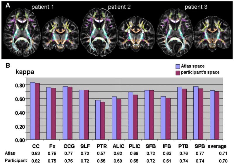
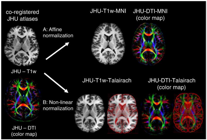
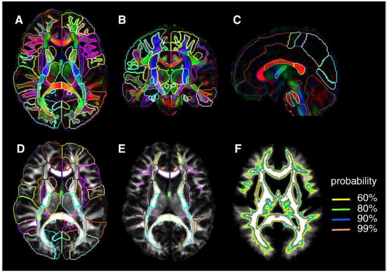
The purpose of this paper is to establish single-participant white matter atlases based on diffusion tensor imaging. As one of the applications of the atlas, automated brain segmentation was performed and the accuracy was measured using Large Deformation Diffeomorphic Metric Mapping (LDDMM). High-quality diffusion tensor imaging (DTI) data from a single-participant were B0-distortion-corrected and transformed to the ICBM-152 atlas or to Talairach coordinates. The deep white matter structures, which have been previously well documented and clearly identified by DTI, were manually segmented. The superficial white matter areas beneath the cortex were defined, based on a population-averaged white matter probability map. The white matter was parcellated into 176 regions based on the anatomical labeling in the ICBM-DTI-81 atlas. The automated parcellation was achieved by warping this parcellation map to normal controls and to Alzheimer's disease patients with severe anatomical atrophy. The parcellation accuracy was measured by a kappa analysis between the automated and manual parcellation at 11 anatomical regions. The kappa values were 0.70 for both normal controls and patients while the inter-rater reproducibility was 0.81 (controls) and 0.82 (patients), suggesting "almost perfect" agreement. A power analysis suggested that the proposed method is suitable for detecting FA and size abnormalities of the white matter in clinical studies.
本文的目的是基于扩散张量成像建立单参与者白质图谱。作为该图谱的应用之一,进行了自动脑分割,并使用大变形微分同胚度量映射(LDDMM)测量了分割精度。对来自单参与者的高质量扩散张量成像(DTI)数据进行B0失真校正,并转换到ICBM - 152图谱或Talairach坐标。对先前已被DTI充分记录并清晰识别的深部白质结构进行手动分割。基于群体平均白质概率图谱定义皮质下方的浅表白质区域。根据ICBM - DTI - 81图谱中的解剖学标记,将白质划分为176个区域。通过将此分割图谱变形到正常对照和患有严重解剖学萎缩的阿尔茨海默病患者,实现自动分割。通过在11个解剖区域对自动分割和手动分割进行kappa分析来测量分割精度。正常对照和患者的kappa值均为0.70,而评分者间的再现性在对照组为0.81,在患者组为0.82,表明具有“几乎完美”的一致性。功效分析表明,所提出的方法适用于在临床研究中检测白质的FA和大小异常。