Russell H. Morgan Department of Radiology and Radiological Science, Division of MR Research, The Johns Hopkins University School of Medicine, Baltimore, MD 21205, USA.
Neuroimage. 2013 Nov 15;82:449-69. doi: 10.1016/j.neuroimage.2013.05.127. Epub 2013 Jun 12.
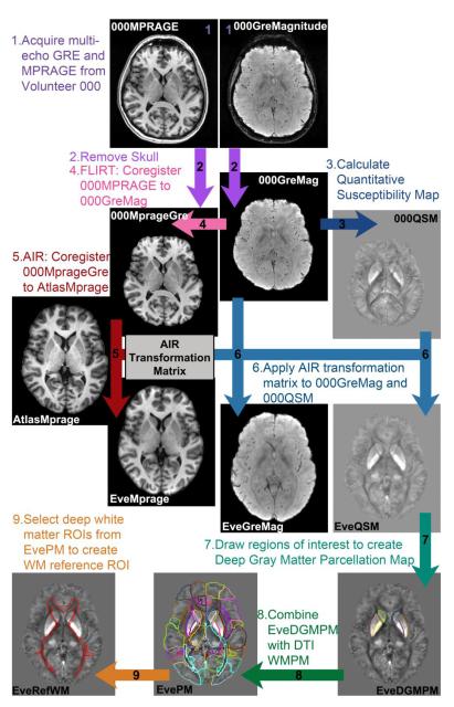
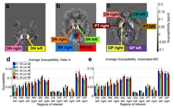
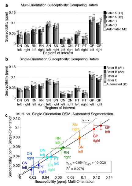
The purpose of this paper is to extend the single-subject Eve atlas from Johns Hopkins University, which currently contains diffusion tensor and T1-weighted anatomical maps, by including contrast based on quantitative susceptibility mapping. The new atlas combines a "deep gray matter parcellation map" (DGMPM) derived from a single-subject quantitative susceptibility map with the previously established "white matter parcellation map" (WMPM) from the same subject's T1-weighted and diffusion tensor imaging data into an MNI coordinate map named the "Everything Parcellation Map in Eve Space," also known as the "EvePM." It allows automated segmentation of gray matter and white matter structures. Quantitative susceptibility maps from five healthy male volunteers (30 to 33 years of age) were coregistered to the Eve Atlas with AIR and Large Deformation Diffeomorphic Metric Mapping (LDDMM), and the transformation matrices were applied to the EvePM to produce automated parcellation in subject space. Parcellation accuracy was measured with a kappa analysis for the left and right structures of six deep gray matter regions. For multi-orientation QSM images, the Kappa statistic was 0.85 between automated and manual segmentation, with the inter-rater reproducibility Kappa being 0.89 for the human raters, suggesting "almost perfect" agreement between all segmentation methods. Segmentation seemed slightly more difficult for human raters on single-orientation QSM images, with the Kappa statistic being 0.88 between automated and manual segmentation, and 0.85 and 0.86 between human raters. Overall, this atlas provides a time-efficient tool for automated coregistration and segmentation of quantitative susceptibility data to analyze many regions of interest. These data were used to establish a baseline for normal magnetic susceptibility measurements for over 60 brain structures of 30- to 33-year-old males. Correlating the average susceptibility with age-based iron concentrations in gray matter structures measured by Hallgren and Sourander (1958) allowed interpolation of the average iron concentration of several deep gray matter regions delineated in the EvePM.
本文旨在扩展约翰霍普金斯大学的单个体 Eve 图谱,该图谱目前包含扩散张量和 T1 加权解剖图谱,并纳入基于定量磁化率映射的对比。新图谱将源自单个体定量磁化率图的“深部灰质分割图谱”(DGMPM)与同一受检者的 T1 加权和扩散张量成像数据的先前建立的“白质分割图谱”(WMPM)结合到一个 MNI 坐标图谱中,称为“Eve 空间中的一切分割图谱”,也称为“EvePM”。它允许自动分割灰质和白质结构。将来自五名健康男性志愿者(30 至 33 岁)的定量磁化率图谱与 AIR 和大变形 diffeomorphic 度量映射(LDDMM)配准到 Eve 图谱中,并将变换矩阵应用于 EvePM 以在受检者空间中产生自动分割。使用左侧和右侧六个深部灰质区域的 kappa 分析测量分割准确性。对于多方位 QSM 图像,自动和手动分割之间的 Kappa 统计量为 0.85,人工评分者的组内可重复性 Kappa 为 0.89,表明所有分割方法之间存在“几乎完美”的一致性。对于单方位 QSM 图像,人工评分者的分割似乎稍微困难一些,自动和手动分割之间的 Kappa 统计量为 0.88,人工评分者之间的 Kappa 统计量为 0.85 和 0.86。总体而言,该图谱为分析许多感兴趣区域的定量磁化率数据的自动配准和分割提供了一种省时的工具。这些数据用于为 30 至 33 岁男性的 60 多个大脑结构建立正常磁化率测量的基线。将平均磁化率与 Hallgren 和 Sourander(1958)测量的灰质结构中铁浓度的年龄相关联,允许对 EvePM 中划定的几个深部灰质区域的平均铁浓度进行插值。