• 文献检索
  • 文档翻译
  • 深度研究
  • 学术资讯
  • Suppr Zotero 插件Zotero 插件
  • 邀请有礼
  • 套餐&价格
  • 历史记录
应用&插件
Suppr Zotero 插件Zotero 插件浏览器插件Mac 客户端Windows 客户端微信小程序
定价
高级版会员购买积分包购买API积分包
服务
文献检索文档翻译深度研究API 文档MCP 服务
关于我们
关于 Suppr公司介绍联系我们用户协议隐私条款
关注我们

Suppr 超能文献

核心技术专利:CN118964589B侵权必究
粤ICP备2023148730 号-1Suppr @ 2026

文献检索

告别复杂PubMed语法,用中文像聊天一样搜索,搜遍4000万医学文献。AI智能推荐,让科研检索更轻松。

立即免费搜索

文件翻译

保留排版,准确专业,支持PDF/Word/PPT等文件格式,支持 12+语言互译。

免费翻译文档

深度研究

AI帮你快速写综述,25分钟生成高质量综述,智能提取关键信息,辅助科研写作。

立即免费体验

通过结构光照明显微镜对活细胞进行超分辨率视频显微镜观察。

Super-resolution video microscopy of live cells by structured illumination.

作者信息

Kner Peter, Chhun Bryant B, Griffis Eric R, Winoto Lukman, Gustafsson Mats G L

机构信息

Department of Biochemistry and Biophysics, San Francisco, California, USA.

出版信息

Nat Methods. 2009 May;6(5):339-42. doi: 10.1038/nmeth.1324. Epub 2009 Apr 26.

DOI:10.1038/nmeth.1324
PMID:19404253
原文链接:https://pmc.ncbi.nlm.nih.gov/articles/PMC2895555/
Abstract

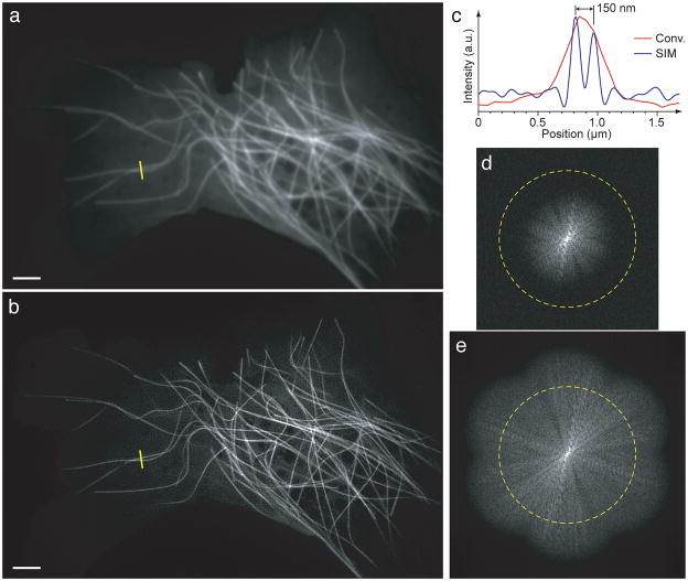
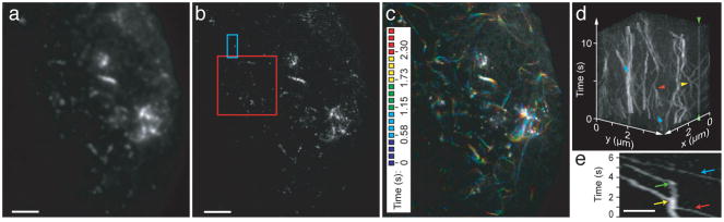
Structured-illumination microscopy can double the resolution of the widefield fluorescence microscope but has previously been too slow for dynamic live imaging. Here we demonstrate a high-speed structured-illumination microscope that is capable of 100-nm resolution at frame rates up to 11 Hz for several hundred time points. We demonstrate the microscope by video imaging of tubulin and kinesin dynamics in living Drosophila melanogaster S2 cells in the total internal reflection mode.

摘要

结构光照明显微镜可将宽场荧光显微镜的分辨率提高一倍,但此前对于动态实时成像来说速度过慢。在此,我们展示了一种高速结构光照明显微镜,它在高达11赫兹的帧率下能够实现100纳米的分辨率,可进行数百个时间点的成像。我们通过在全内反射模式下对活的黑腹果蝇S2细胞中的微管蛋白和驱动蛋白动力学进行视频成像,展示了该显微镜。

https://cdn.ncbi.nlm.nih.gov/pmc/blobs/7d2c/2895555/d03dd26af99a/nihms105932f3.jpg
https://cdn.ncbi.nlm.nih.gov/pmc/blobs/7d2c/2895555/aee7da1e3e74/nihms105932f1.jpg
https://cdn.ncbi.nlm.nih.gov/pmc/blobs/7d2c/2895555/ea1a78935384/nihms105932f2.jpg
https://cdn.ncbi.nlm.nih.gov/pmc/blobs/7d2c/2895555/d03dd26af99a/nihms105932f3.jpg
https://cdn.ncbi.nlm.nih.gov/pmc/blobs/7d2c/2895555/aee7da1e3e74/nihms105932f1.jpg
https://cdn.ncbi.nlm.nih.gov/pmc/blobs/7d2c/2895555/ea1a78935384/nihms105932f2.jpg
https://cdn.ncbi.nlm.nih.gov/pmc/blobs/7d2c/2895555/d03dd26af99a/nihms105932f3.jpg

相似文献

1
Super-resolution video microscopy of live cells by structured illumination.通过结构光照明显微镜对活细胞进行超分辨率视频显微镜观察。
Nat Methods. 2009 May;6(5):339-42. doi: 10.1038/nmeth.1324. Epub 2009 Apr 26.
2
Live, video-rate super-resolution microscopy using structured illumination and rapid GPU-based parallel processing.使用结构光照和基于 GPU 的快速并行处理的实时、视频速率超分辨率显微镜。
Microsc Microanal. 2011 Apr;17(2):191-6. doi: 10.1017/S1431927611000158. Epub 2011 Mar 9.
3
Dynamics of myosin, microtubules, and Kinesin-6 at the cortex during cytokinesis in Drosophila S2 cells.果蝇S2细胞胞质分裂期间皮层中肌球蛋白、微管和驱动蛋白-6的动态变化
J Cell Biol. 2009 Sep 7;186(5):727-38. doi: 10.1083/jcb.200902083. Epub 2009 Aug 31.
4
Super-resolution 3D microscopy of live whole cells using structured illumination.使用结构光照明显微镜对活的整个细胞进行超高分辨率 3D 成像。
Nat Methods. 2011 Oct 16;8(12):1044-6. doi: 10.1038/nmeth.1734.
5
Microtubules and microscopes: how the development of light microscopic imaging technologies has contributed to discoveries about microtubule dynamics in living cells.微管与显微镜:光学显微镜成像技术的发展如何助力关于活细胞中微管动力学的发现。
Mol Biol Cell. 1998 Dec;9(12):3263-71. doi: 10.1091/mbc.9.12.3263.
6
Video-rate multi-color structured illumination microscopy with simultaneous real-time reconstruction.视频速率多色结构光照明显微镜,具有实时同步重建功能。
Nat Commun. 2019 Sep 20;10(1):4315. doi: 10.1038/s41467-019-12165-x.
7
Fast optical sectioning obtained by structured illumination microscopy using a digital mirror device.基于数字微镜器件的结构光照明显微镜快速光学切片。
J Biomed Opt. 2013 Jun;18(6):060503. doi: 10.1117/1.JBO.18.6.060503.
8
Recent advancements in structured-illumination microscopy toward live-cell imaging.结构照明显微镜在活细胞成像方面的最新进展。
Microscopy (Oxf). 2015 Aug;64(4):237-49. doi: 10.1093/jmicro/dfv034. Epub 2015 Jun 30.
9
Widefield and total internal reflection fluorescent structured illumination microscopy with scanning galvo mirrors.宽场和全内反射荧光结构光照明显微镜与扫描振镜。
J Biomed Opt. 2018 Apr;23(4):1-9. doi: 10.1117/1.JBO.23.4.046007.
10
A novel microtubule nucleation pathway for meiotic spindle assembly in oocytes.一种新的微管成核途径,用于卵母细胞减数分裂纺锤体的组装。
J Cell Biol. 2018 Oct 1;217(10):3431-3445. doi: 10.1083/jcb.201803172. Epub 2018 Aug 7.

引用本文的文献

1
Advances in High-Speed Structured Illumination Microscopy.高速结构光照明显微镜技术的进展
Front Phys. 2021 May;9. doi: 10.3389/fphy.2021.672555. Epub 2021 May 28.
2
Cellular optical imaging techniques: a dynamic advancing frontier.细胞光学成像技术:一个动态发展的前沿领域。
Sci China Life Sci. 2025 Jul 16. doi: 10.1007/s11427-024-2916-5.
3
Projective oblique plane structured illumination microscopy.投影斜平面结构照明显微镜

本文引用的文献

1
Kinesin-5-dependent poleward flux and spindle length control in Drosophila embryo mitosis.果蝇胚胎有丝分裂中驱动蛋白5依赖性的极向通量与纺锤体长度控制
Mol Biol Cell. 2009 Mar;20(6):1749-62. doi: 10.1091/mbc.e08-10-1033. Epub 2009 Jan 21.
2
Sub-100-nanometre resolution in total internal reflection fluorescence microscopy.全内反射荧光显微镜中的亚100纳米分辨率
J Microsc. 2008 Oct;232(1):99-105. doi: 10.1111/j.1365-2818.2008.02075.x.
3
Structured illumination in total internal reflection fluorescence microscopy using a spatial light modulator.
Npj Imaging. 2023 Nov 28;1(1):2. doi: 10.1038/s44303-023-00002-2.
4
Advancing Multicolor Super-Resolution Volume Imaging: Illuminating Complex Cellular Dynamics.推进多色超分辨率体积成像:揭示复杂的细胞动力学。
JACS Au. 2025 Jun 9;5(6):2388-2419. doi: 10.1021/jacsau.5c00314. eCollection 2025 Jun 23.
5
Two-Dimensional Nonlinear Structured Illumination Microscopy with rsEGFP2.基于rsEGFP2的二维非线性结构照明显微术
bioRxiv. 2025 May 15:2025.05.11.653285. doi: 10.1101/2025.05.11.653285.
6
Physical twinning for joint encoding-decoding optimization in computational optics: a review.计算光学中用于联合编码-解码优化的物理孪生:综述
Light Sci Appl. 2025 Apr 15;14(1):162. doi: 10.1038/s41377-025-01810-4.
7
LED-based multicolor extended resolution transmission fluorescence microscopy.基于发光二极管的多色扩展分辨率透射荧光显微镜。
J Biomed Opt. 2025 Apr;30(4):046501. doi: 10.1117/1.JBO.30.4.046501. Epub 2025 Apr 8.
8
Super-resolution microscopy based on the inherent fluctuations of dye molecules.基于染料分子固有涨落的超分辨率显微镜技术。
Biomed Opt Express. 2025 Feb 4;16(3):910-921. doi: 10.1364/BOE.533263. eCollection 2025 Mar 1.
9
Fluorescent labeling strategies for molecular bioimaging.用于分子生物成像的荧光标记策略。
Biophys Rep (N Y). 2025 Mar 12;5(1):100200. doi: 10.1016/j.bpr.2025.100200. Epub 2025 Feb 12.
10
Elucidating subcellular architecture and dynamics at isotropic 100-nm resolution with 4Pi-SIM.利用4Pi-SIM以各向同性100纳米分辨率阐明亚细胞结构和动力学。
Nat Methods. 2025 Feb;22(2):335-347. doi: 10.1038/s41592-024-02515-z. Epub 2024 Dec 23.
使用空间光调制器的全内反射荧光显微镜中的结构照明
Opt Lett. 2008 Jul 15;33(14):1629-31. doi: 10.1364/ol.33.001629.
4
Augmin: a protein complex required for centrosome-independent microtubule generation within the spindle.Augmin:一种纺锤体内不依赖中心体的微管生成所需的蛋白质复合体。
J Cell Biol. 2008 May 5;181(3):421-9. doi: 10.1083/jcb.200711053. Epub 2008 Apr 28.
5
Live-cell photoactivated localization microscopy of nanoscale adhesion dynamics.纳米级黏附动力学的活细胞光活化定位显微镜技术
Nat Methods. 2008 May;5(5):417-23. doi: 10.1038/nmeth.1202. Epub 2008 Apr 13.
6
Three-dimensional resolution doubling in wide-field fluorescence microscopy by structured illumination.通过结构光照实现宽场荧光显微镜三维分辨率翻倍
Biophys J. 2008 Jun;94(12):4957-70. doi: 10.1529/biophysj.107.120345. Epub 2008 Mar 7.
7
Video-rate far-field optical nanoscopy dissects synaptic vesicle movement.视频速率远场光学纳米显微镜解析突触小泡运动。
Science. 2008 Apr 11;320(5873):246-9. doi: 10.1126/science.1154228. Epub 2008 Feb 21.
8
Spindly, a novel protein essential for silencing the spindle assembly checkpoint, recruits dynein to the kinetochore.Spindly是一种对纺锤体组装检查点沉默至关重要的新型蛋白质,它将动力蛋白招募到动粒。
J Cell Biol. 2007 Jun 18;177(6):1005-15. doi: 10.1083/jcb.200702062.
9
Poleward tubulin flux in spindles: regulation and function in mitotic cells.纺锤体中微管蛋白向极运输:有丝分裂细胞中的调控与功能
Mol Biol Cell. 2007 Aug;18(8):3094-104. doi: 10.1091/mbc.e06-11-0994. Epub 2007 Jun 6.
10
Two-dimensional standing wave total internal reflection fluorescence microscopy: superresolution imaging of single molecular and biological specimens.二维驻波全内反射荧光显微镜:单分子和生物样本的超分辨率成像
Biophys J. 2007 Sep 1;93(5):1747-57. doi: 10.1529/biophysj.106.097907. Epub 2007 May 4.