Wu Yang, Nelson Morag M, Quaile Andrew, Xia Dong, Wastling Jonathan M, Craig Alister
Liverpool School of Tropical Medicine, Pembroke Place, Liverpool L3 5QA, UK.
Malar J. 2009 May 18;8:105. doi: 10.1186/1475-2875-8-105.
Previous comparative proteomic analysis on Plasmodium falciparum isolates of different adhesion properties suggested that protein phosphorylation varies between isolates with different cytoadherence properties. But the extent and dynamic changes in phosphorylation have not been systematically studied. As a baseline for these future studies, this paper examined changes in the phosphoproteome of parasitized red blood cells (pRBC).
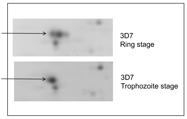
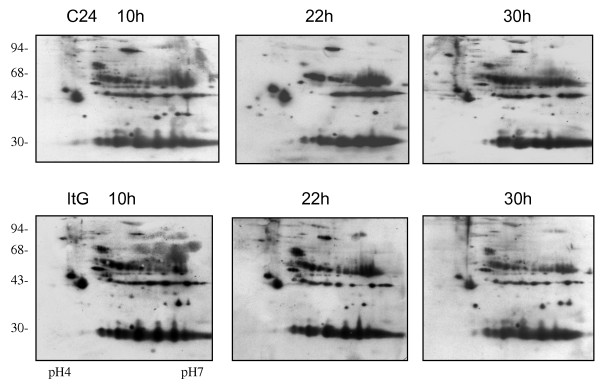
Metabolic labelling with [35S] methionine on pRBC and 2D gel electrophoresis (2-DE) has previously been used to show the expression of parasite proteins and changes in protein iso-electric point (PI). 2-DE of different parasite strains was combined with immunoblotting using monoclonal antibodies specifically to phosphorylated serine/threonine and tyrosine, to obtain the phosphorylation profiles throughout the erythrocytic lifecycle. Affinity chromatography was used to purify/enrich phosphorylated proteins and these proteins from mature trophozoite stages which were identified using high-accuracy mass spectrometry and MASCOT search.
2D-immunoblots showed that P. falciparum infection greatly increased phosphorylation of a set of proteins in pRBC, the dominant size classes for phosphorylated tyrosine proteins were 95, 60, 50 and 30 kDa and for phosphorylated serine/threonine were 120, 95, 60, 50, 43, 40 and 30 kDa. The most abundant molecules from 2D-gel mapping of phosphorylated proteins in ItG infected RBCs were identified by MALDI-TOF. A proteomic overview of phosphorylated proteins in pRBC was achieved by using complementary phosphorylated protein enrichment techniques combined with nano-flow LC/MS/MS analysis and MASCOT MS/MS ions search with phosphorylation as variable modifications. The definite phosphoproteins of pRBC are reported and discussed.
Protein phosphorylation is a major process in P. falciparum-parasitized erythrocytes. Preliminary screens identified 170 P. falciparum proteins and 77 human proteins as phosphorylated protein in pRBC, while only 48 human proteins were identified in the corresponding fractions from uninfected RBC. Refinement of the search to include significant ion scores indicating a specific phospho-peptide identified 21 P. falciparum proteins and 14 human proteins from pRBC, 13 host proteins were identified from normal RBC. The results achieved by complementary techniques consistently reflect a reliable proteomic overview of pRBC.
先前对具有不同黏附特性的恶性疟原虫分离株进行的比较蛋白质组学分析表明,蛋白质磷酸化在具有不同细胞黏附特性的分离株之间存在差异。但磷酸化的程度和动态变化尚未得到系统研究。作为这些未来研究的基线,本文研究了被寄生红细胞(pRBC)磷酸化蛋白质组的变化。
先前已使用[35S]甲硫氨酸对pRBC进行代谢标记和二维凝胶电泳(2-DE)来显示寄生虫蛋白的表达以及蛋白质等电点(PI)的变化。将不同寄生虫菌株的2-DE与使用特异性针对磷酸化丝氨酸/苏氨酸和酪氨酸的单克隆抗体的免疫印迹相结合,以获得整个红细胞生命周期的磷酸化图谱。亲和色谱法用于纯化/富集磷酸化蛋白质,并用高精度质谱和MASCOT搜索鉴定来自成熟滋养体阶段的这些蛋白质。
二维免疫印迹显示,恶性疟原虫感染极大地增加了pRBC中一组蛋白质的磷酸化,磷酸化酪氨酸蛋白的主要大小类别为95、60、50和30 kDa,磷酸化丝氨酸/苏氨酸蛋白的主要大小类别为120、95、60、50、43、40和30 kDa。通过基质辅助激光解吸电离飞行时间质谱(MALDI-TOF)鉴定了感染ItG的红细胞中磷酸化蛋白质二维凝胶图谱中最丰富的分子。通过使用互补的磷酸化蛋白质富集技术、纳流液相色谱/串联质谱(nano-flow LC/MS/MS)分析以及以磷酸化为可变修饰的MASCOT MS/MS离子搜索,实现了对pRBC中磷酸化蛋白质的蛋白质组学概述。报道并讨论了pRBC中确定的磷酸化蛋白质。
蛋白质磷酸化是恶性疟原虫寄生红细胞中的一个主要过程。初步筛选确定了170种恶性疟原虫蛋白和77种人类蛋白为pRBC中的磷酸化蛋白,而在未感染红细胞的相应组分中仅鉴定出48种人类蛋白。将搜索范围细化以纳入表明特定磷酸肽的显著离子得分,从pRBC中鉴定出21种恶性疟原虫蛋白和14种人类蛋白,从正常红细胞中鉴定出13种宿主蛋白。通过互补技术获得的结果一致反映了pRBC可靠的蛋白质组学概述。