Yang Dongli, Elner Susan G, Lin Li-Ren, Reddy Venkat N, Petty Howard R, Elner Victor M
Department of Ophthalmology and Visual Sciences, University of Michigan, Ann Arbor, Michigan 48105-0714, USA.
Invest Ophthalmol Vis Sci. 2009 Oct;50(10):4998-5005. doi: 10.1167/iovs.09-3620. Epub 2009 May 20.
Oxidative stress of the retinal pigment epithelium by reactive oxygen species and monocytic infiltration have been implicated in age-related macular degeneration. The purpose of this study was to determine the role of superoxide anions (O(2)(-)) in mononuclear phagocyte-induced RPE apoptosis.
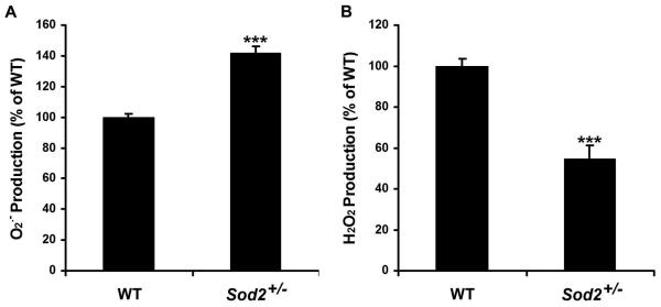
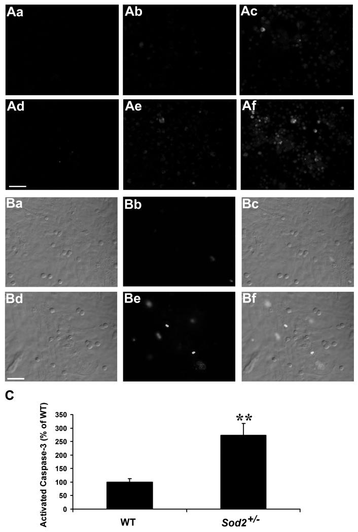
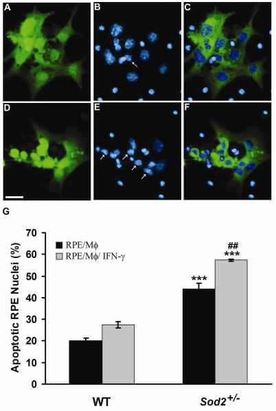
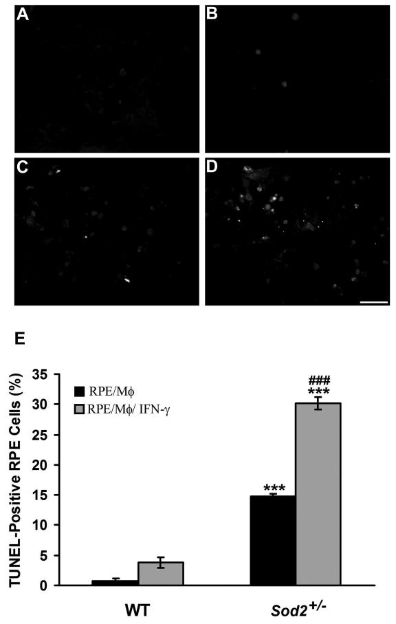
Mouse RPE cell cultures were established from wild-type and heterozygous superoxide dismutase 2-knockout (Sod2(+/-)) mice. The intracellular reactive oxygen species, O(2)(-) and hydrogen peroxide, were measured by using dihydroethidium assay and 5-(and 6)-chloromethyl-2',7'-dichlorodihydrofluorescence diacetate, acetyl ester assay, respectively. RPE apoptosis was evaluated by Hoechst staining and terminal deoxynucleotidyltransferase dUTP nick-end labeling assay. Changes in mitochondrial membrane potential were detected by 5,5',6,6'-tetrachloro-1,1',3,3'-tetraethylbenzimidazolylcarbocyanine iodide dye. Activated caspases and caspase-3 were detected in situ by FITC-VAD-fmk staining and caspase-3 substrate, respectively.
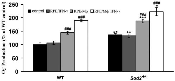
Mononuclear phagocytes and interferon-gamma-activated mononuclear phagocytes induced the production of intracellular RPE O(2)(-), a decrease in RPE mitochondrial membrane potential, caspase activation, and apoptosis of mouse RPE cells. All theses changes were significantly enhanced in the Sod2(+/-) RPE cells. Activated mononuclear phagocytes induced more of these oxidative and apoptotic changes in RPE cells than did unstimulated mononuclear phagocytes.
The authors have shown that the decreased expression of SOD2 and increased superoxide production correlate with RPE apoptosis induced by unstimulated and activated mononuclear phagocytes. The authors suggest that elevated O(2)(-) levels due to genetic abnormalities of SOD2 or immunologic activation of mononuclear phagocytes lead to greater levels of RPE apoptosis. The present study could serve as a useful model to characterize RPE/phagocyte interaction in AMD and other retinal diseases.
活性氧导致的视网膜色素上皮氧化应激和单核细胞浸润与年龄相关性黄斑变性有关。本研究的目的是确定超氧阴离子(O₂⁻)在单核吞噬细胞诱导的视网膜色素上皮细胞凋亡中的作用。
从小鼠野生型和杂合子超氧化物歧化酶2基因敲除(Sod2⁺/⁻)小鼠建立视网膜色素上皮细胞培养。分别使用二氢乙锭检测法和5-(及6)-氯甲基-2′,7′-二氯二氢荧光素二乙酸酯、乙酰酯检测法测量细胞内活性氧、O₂⁻和过氧化氢。通过Hoechst染色和末端脱氧核苷酸转移酶dUTP缺口末端标记检测法评估视网膜色素上皮细胞凋亡。用5,5′,6,6′-四氯-1,1′,3,3′-四乙基苯并咪唑基羰花青碘化物染料检测线粒体膜电位的变化。分别通过FITC-VAD-fmk染色和半胱天冬酶-3底物原位检测活化的半胱天冬酶和半胱天冬酶-3。
单核吞噬细胞和干扰素-γ激活的单核吞噬细胞诱导小鼠视网膜色素上皮细胞产生细胞内O₂⁻,导致视网膜色素上皮线粒体膜电位降低、半胱天冬酶激活和细胞凋亡。所有这些变化在Sod2⁺/⁻视网膜色素上皮细胞中显著增强。与未刺激的单核吞噬细胞相比,活化的单核吞噬细胞在视网膜色素上皮细胞中诱导更多的这些氧化和凋亡变化。
作者表明,超氧化物歧化酶2表达降低和超氧化物产生增加与未刺激和活化的单核吞噬细胞诱导的视网膜色素上皮细胞凋亡相关。作者认为,由于超氧化物歧化酶2的基因异常或单核吞噬细胞的免疫激活导致O₂⁻水平升高,从而导致更高水平的视网膜色素上皮细胞凋亡。本研究可作为一个有用的模型,用于表征年龄相关性黄斑变性和其他视网膜疾病中视网膜色素上皮/吞噬细胞的相互作用。