Lee Jun Hyuck, Rangarajan Erumbi S, Yogesha S D, Izard Tina
Cell Adhesion Laboratory, Department of Cancer Biology, The Scripps Research Institute, Jupiter, FL 33458, USA.
Structure. 2009 Jun 10;17(6):833-42. doi: 10.1016/j.str.2009.04.010.
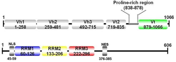
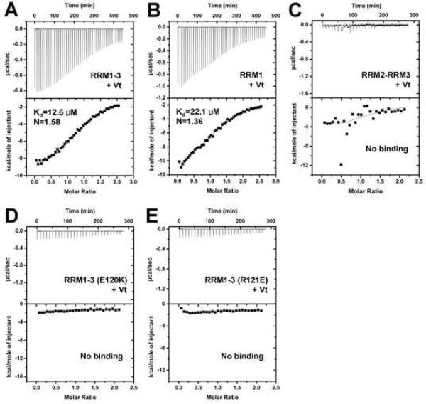
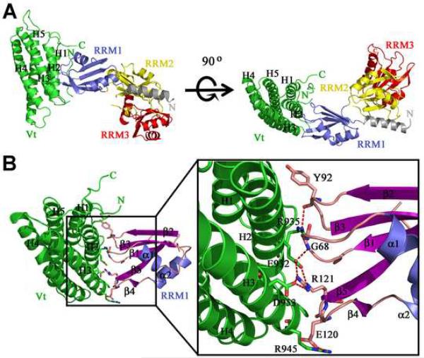
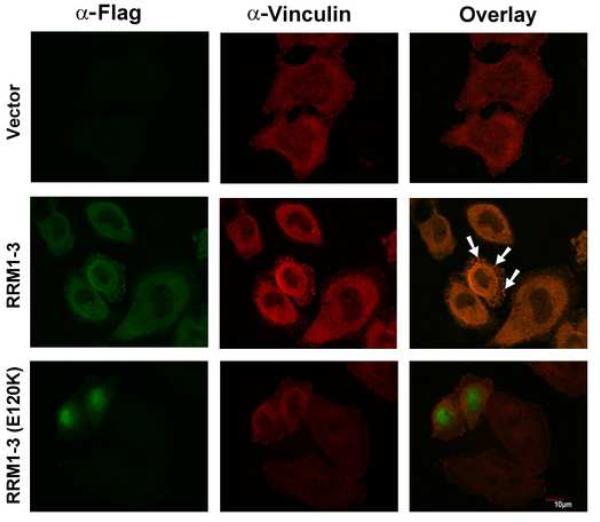
The translational machinery of the cell relocalizes to focal adhesions following the activation of integrin receptors. This response allows for rapid, local production of components needed for adhesion complex assembly and signaling. Vinculin links focal adhesions to the actin cytoskeleton following its activation by integrin signaling, which severs intramolecular interactions of vinculin's head and tail (Vt) domains. Our vinculin:raver1 crystal structures and binding studies show that activated Vt selectively interacts with one of the three RNA recognition motifs of raver1, that the vinculin:raver1 complex binds to F-actin, and that raver1 binds selectively to RNA, including a sequence found in vinculin mRNA. Further, mutation of residues that mediate interaction of raver1 with vinculin abolish their colocalization in cells. These findings suggest a feed-forward model where vinculin activation at focal adhesions provides a scaffold for recruitment of raver1 and its mRNA cargo to facilitate the production of components of adhesion complexes.
整合素受体激活后,细胞的翻译机制会重新定位于粘着斑。这种反应使得粘着斑组装和信号传导所需的成分能够快速在局部产生。纽蛋白在整合素信号激活后将粘着斑与肌动蛋白细胞骨架相连,这会切断纽蛋白头部和尾部(Vt)结构域的分子内相互作用。我们的纽蛋白:raver1晶体结构和结合研究表明,活化的Vt选择性地与raver1的三个RNA识别基序之一相互作用,纽蛋白:raver1复合物与F-肌动蛋白结合,并且raver1选择性地与RNA结合,包括在纽蛋白mRNA中发现的一个序列。此外,介导raver1与纽蛋白相互作用的残基发生突变会消除它们在细胞中的共定位。这些发现提示了一种前馈模型,即粘着斑处的纽蛋白激活为raver1及其mRNA货物的募集提供了一个支架,以促进粘着斑复合物成分的产生。