Soul Janet S, Robertson Richard L, Wypij David, Bellinger David C, Visconti Karen J, du Plessis Adré J, Kussman Barry D, Scoppettuolo Lisa A, Pigula Frank, Jonas Richard A, Newburger Jane W
Department of Neurology, Children's Hospital Boston, Boston, MA 02115, USA.
J Thorac Cardiovasc Surg. 2009 Aug;138(2):374-81. doi: 10.1016/j.jtcvs.2009.02.027. Epub 2009 Apr 10.
Perioperative stroke and periventricular leukomalacia have been reported to occur commonly in infants with congenital heart disease. We aimed to determine the incidence and type of brain injury in infants undergoing 2-ventricle repair in infancy and to determine risk factors associated with such injury.
Forty-eight infants enrolled in a trial comparing 2 different hematocrits during surgical repair of congenital heart disease underwent brain magnetic resonance imaging scans and neurodevelopmental testing at 1 year of age.
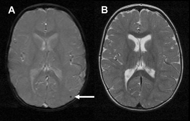
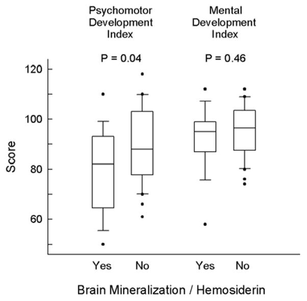
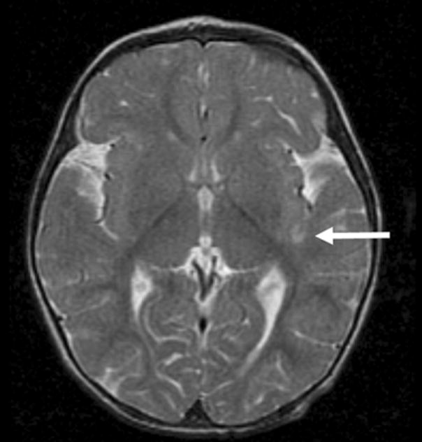
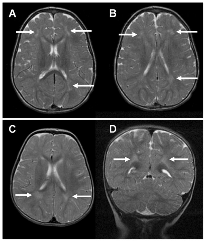
Eighteen (38%) of our subjects had tiny foci of hemosiderin by susceptibility imaging, without evidence of abnormalities in corresponding regions on conventional magnetic resonance imaging sequences. Subjects with foci of hemosiderin had a significantly lower Psychomotor Developmental Index at 1 year of age (79.6 +/- 16.5, mean +/- standard deviation) compared with subjects without these foci (89.5 +/- 15.3; P = .04). Older age at surgery and diagnostic group were significantly associated with the presence of hemosiderin foci. Only 1 subject had a small stroke (2%), and 2 subjects had periventricular leukomalacia (4%).
Foci of hemosiderin without radiologic evidence of ischemic brain injury are an abnormality associated with adverse neurodevelopmental outcome not previously described in magnetic resonance imaging studies of children with surgically repaired congenital heart disease. The association of hemosiderin foci with older age at surgery and cardiac diagnosis, and not with risk factors associated with brain injury, in previous studies suggests that the cause and pathogenesis of this abnormality are different from ischemic brain lesions reported previously.
据报道,围手术期卒中及脑室周围白质软化症在先天性心脏病婴儿中很常见。我们旨在确定婴儿期接受双心室修复的婴儿脑损伤的发生率和类型,并确定与此类损伤相关的危险因素。
48名参加先天性心脏病手术修复期间比较两种不同血细胞比容试验的婴儿在1岁时接受了脑磁共振成像扫描和神经发育测试。
通过敏感性成像,我们的18名(38%)受试者有微小的含铁血黄素病灶,而在传统磁共振成像序列上相应区域无异常证据。有含铁血黄素病灶的受试者在1岁时的精神运动发育指数(平均±标准差,79.6±16.5)显著低于无这些病灶的受试者(89.5±15.3;P = 0.04)。手术时年龄较大和诊断组与含铁血黄素病灶的存在显著相关。只有1名受试者发生小卒中(2%),2名受试者发生脑室周围白质软化症(4%)。
无缺血性脑损伤放射学证据的含铁血黄素病灶是一种与不良神经发育结局相关的异常情况,此前在接受手术修复的先天性心脏病儿童的磁共振成像研究中未描述过。在先前的研究中,含铁血黄素病灶与手术时年龄较大和心脏诊断相关,而与脑损伤相关危险因素无关,这表明这种异常情况的病因和发病机制与先前报道的缺血性脑病变不同。