Zimmermann P, Dietrich T, Bock F, Horn F K, Hofmann-Rummelt C, Kruse F E, Cursiefen C
Department of Ophthalmology, University of Erlangen-Nürnberg, Schwabachanlage 6, 91054 Erlangen, Germany.
Br J Ophthalmol. 2009 Nov;93(11):1529-34. doi: 10.1136/bjo.2008.147355. Epub 2009 Jul 23.
To evaluate whether tumour-associated lymphangiogenesis, that is the formation of new lymphatic vessels (LVs) induced by a tumour, occurs in and around conjunctival malignant melanoma (MM).
Clinical files and conjunctival specimens of 20 patients with histologically diagnosed conjunctival MM were analysed. Sections were stained with LYVE-1 and podoplanin antibodies as specific lymphatic endothelial markers and Ki67 as proliferation marker. The tumour area and the area covered by LV (LVA), LV number (LVN) and LV density (LVD) were measured within the tumour and in the peritumoural area in digital images of the specimen. The LV results were correlated with the histopathological characteristics, tumour location, recurrence rate, mitomycin C therapy and presence of metastases.
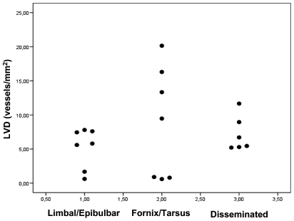
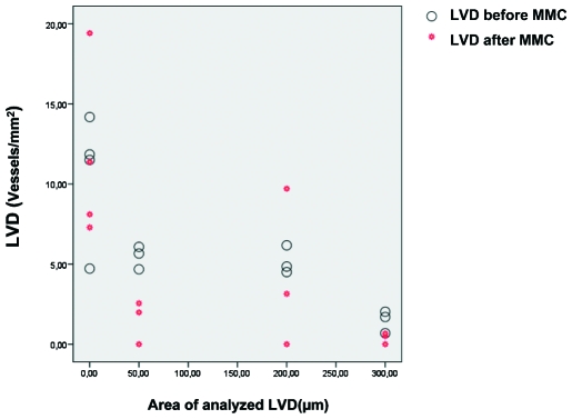
LVs were detected in all specimens within the tumour and peritumourally. Significantly more Ki67(+) proliferating lymphatic endothelial cells were detected in the tumour and in the peritumoural tissue up to 300 microm compared with the surrounding normal conjunctiva (>300 microm distance). There was a slightly positive correlation between the tumour size and the LVN and LVA in the 50 microm zone adjacent to the tumour. We did not find any significant correlations between LVs and histopathological and clinical characteristics (location, shape, relapses, metastases), possibly due to the small sample sizes. Non-limbal tumours with involvement of tarsus or fornix showed a tendency towards a higher LVD compared with limbal tumours.
Conjunctival MMs display tumour-associated LV within and around the tumour. The MM seems to induce lymphangiogenesis not only in the tumour, but also in its proximity.
评估肿瘤相关淋巴管生成,即由肿瘤诱导的新淋巴管形成,是否发生于结膜恶性黑色素瘤(MM)及其周围。
分析20例经组织学诊断为结膜MM患者的临床资料和结膜标本。切片用LYVE-1和血小板内皮细胞黏附分子抗体作为特异性淋巴管内皮标记物,用Ki67作为增殖标记物进行染色。在标本的数字图像中测量肿瘤内和肿瘤周围区域的肿瘤面积、淋巴管覆盖面积(LVA)、淋巴管数量(LVN)和淋巴管密度(LVD)。将淋巴管结果与组织病理学特征、肿瘤位置、复发率、丝裂霉素C治疗及转移情况进行关联分析。
在所有标本的肿瘤内及肿瘤周围均检测到淋巴管。与周围正常结膜(距离>300微米)相比,在肿瘤内及距离肿瘤300微米以内的肿瘤周围组织中检测到显著更多的Ki67(+)增殖淋巴管内皮细胞。在与肿瘤相邻的50微米区域内,肿瘤大小与LVN和LVA之间存在轻度正相关。我们未发现淋巴管与组织病理学及临床特征(位置、形状、复发、转移)之间存在任何显著相关性,可能是由于样本量较小。与角膜缘肿瘤相比,累及睑板或穹窿的非角膜缘肿瘤显示出LVD较高的趋势。
结膜MM在肿瘤内及周围显示出肿瘤相关淋巴管。MM似乎不仅在肿瘤内,而且在其附近诱导淋巴管生成。