Institute of Neurology, University College London, Queen Square, London, UK.
Neuroimage. 2010 Jan 1;49(1):185-92. doi: 10.1016/j.neuroimage.2009.07.037. Epub 2009 Jul 23.
Loss of brain volume in first-episode psychosis can be detected using conventional magnetic resonance imaging (MRI), but subtle changes--not leading to reduction in volume--that may contribute to clinical and cognitive abnormalities, may go undetected. Magnetization transfer imaging (MTI), a technique more sensitive to subtle neuropathological changes than conventional MRI, could yield important information on the extent and nature of structural abnormalities.
Forty-eight patients (33 males) from a population-based sample with first-episode psychosis (41 with schizophrenia and 7 with schizoaffective psychosis) and 47 healthy volunteers (27 males) were studied. Differences in magnetization transfer ratio (MTR) and white and grey matter volumes between groups were investigated.
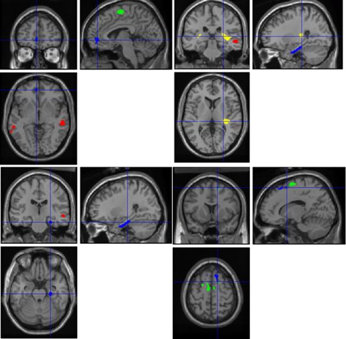
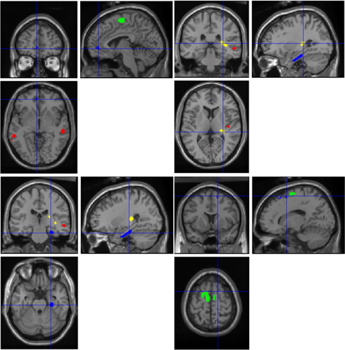
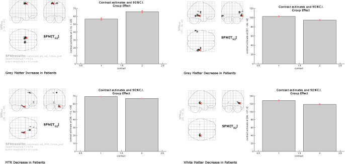
In patients, MTR was reduced in right entorhinal cortex, fusiform, dentate and superior frontal gyri and in left superior frontal and inferior/rostral cingulate gyri. Grey matter volume was reduced in right insula, frontal operculum and middle and superior temporal gyri and in left middle temporal gyrus. Grey matter volume increases were seen in patients in the superior frontal gyrus. White matter volume loss was found adjacent to grey matter loss. In patients MTR was lower in all areas of volumetric differences between groups suggesting that both changes may be related. Similar findings were observed when patients with schizoaffective psychosis were removed from the analysis. The correlations between clinical and MRI parameters did not survive correction for multiple comparisons.
MTI frontal and temporal abnormalities suggesting neuroaxonal and myelin changes were more extensive in our patients than those detected with conventional MRI. Our findings also suggest that there is regional variation in the severity of structural brain abnormalities.
首次发作精神分裂症患者的脑容量损失可通过常规磁共振成像(MRI)检测到,但可能导致临床和认知异常的细微变化(不会导致体积减少)可能未被发现。磁化传递成像(MTI)是一种比常规 MRI 更敏感的检测细微神经病理学变化的技术,它可以提供有关结构异常的范围和性质的重要信息。
对来自基于人群的首次发作精神分裂症患者(41 例精神分裂症和 7 例分裂情感性精神病)和 47 名健康志愿者(27 名男性)的 48 名患者(33 名男性)进行了研究。研究了组间磁化传递率(MTR)和白质及灰质体积的差异。
在患者中,右侧内嗅皮质、梭状回、齿状回和额上回以及左侧额上回和额下回/前扣带回的 MTR 降低。灰质体积减少见于右侧岛叶、额眶回和中颞回以及左侧中颞回。患者的额上回灰质体积增加。在灰质丢失的部位发现了白质体积的丢失。在患者中,所有组间体积差异的 MTR 均较低,表明这两种变化可能相关。当从分析中去除分裂情感性精神病患者时,也观察到了类似的发现。临床和 MRI 参数之间的相关性在经过多次比较校正后并未保留。
MTI 额叶和颞叶异常提示神经轴突和髓鞘变化在我们的患者中比常规 MRI 检测到的更为广泛。我们的研究结果还表明,结构脑异常的严重程度存在区域性差异。