Chewning Joseph H, Dugger Kari J, Chaudhuri Tandra R, Zinn Kurt R, Weaver Casey T
Department of Pediatrics, University of Alabama at Birmingham, Birmingham, Alabama 35294, USA.
BMC Immunol. 2009 Aug 3;10:44. doi: 10.1186/1471-2172-10-44.
Rapid clonal expansion of T cells occurs in response to antigenic challenges. The kinetics of the T cell response has previously been described using tissue-based studies performed at defined time points. Luciferase bioluminescence has recently been utilized for non-invasive analysis of in vivo biologic processes in real-time.
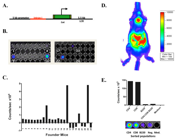
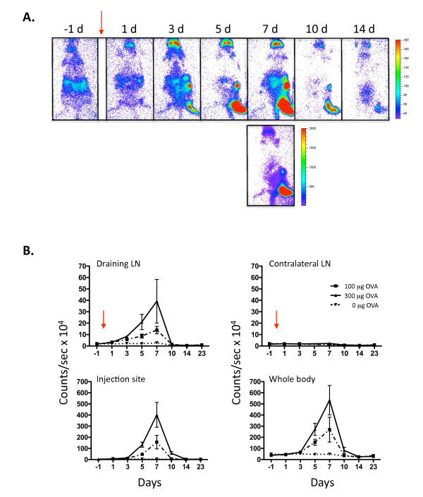
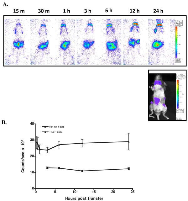
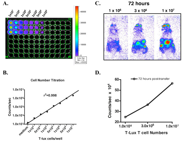
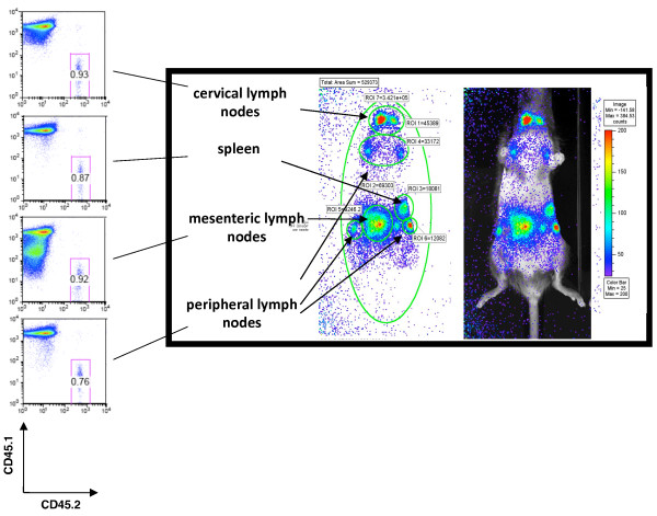
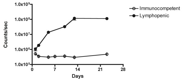
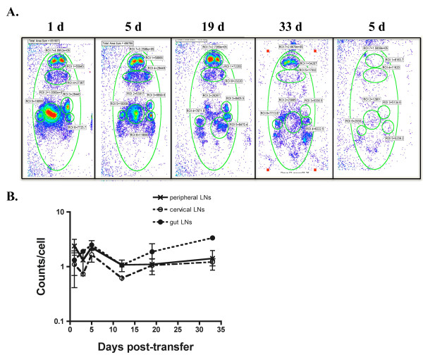
We have created a novel transgenic mouse model (T-Lux) using a human CD2 mini-gene to direct luciferase expression specifically to the T cell compartment. T-Lux T cells demonstrated normal homing patterns within the intact mouse and following adoptive transfer. Bioluminescent signal correlated with T cell numbers in the whole body images as well as within specific organ regions of interest. Following transfer into lymphopenic (RAG2-/-) recipients, homeostatic proliferation of T-Lux T cells was visualized using bioluminescent imaging. Real-time bioluminescent analysis of CD4+ T cell antigen-specific responses enabled real-time comparison of the kinetics and magnitude of clonal expansion and contraction in the inductive lymph node and tissue site of antigen injection. T cell expansion was dose-dependent despite the presence of supraphysiologic numbers of OVA-specific OT-II transgenic TCR T-Lux T cells. CD4+ T cells subsequently underwent a rapid (3-4 day) contraction phase in the draining lymph node, with a delayed contraction in the antigen delivery site, with bioluminescent signal diminished below initial levels, representing TCR clonal frequency control.
The T-Lux mouse provides a novel, efficient model for tracking in vivo aspects of the CD4+ T cell response to antigen, providing an attractive approach for studies directed at immunotherapy or vaccine design.
T细胞会因抗原刺激而发生快速克隆扩增。此前,T细胞反应的动力学已通过在特定时间点进行的基于组织的研究来描述。荧光素酶生物发光最近已被用于实时非侵入性分析体内生物学过程。
我们利用人CD2微型基因创建了一种新型转基因小鼠模型(T-Lux),以将荧光素酶表达特异性地导向T细胞区室。T-Lux T细胞在完整小鼠体内以及过继转移后表现出正常的归巢模式。生物发光信号与全身图像以及特定感兴趣器官区域内的T细胞数量相关。将T-Lux T细胞转移到淋巴细胞减少的(RAG2-/-)受体小鼠后,利用生物发光成像观察到了T-Lux T细胞的稳态增殖。对CD4+ T细胞抗原特异性反应的实时生物发光分析能够实时比较诱导淋巴结和抗原注射组织部位中克隆扩增和收缩的动力学及幅度。尽管存在超生理数量的OVA特异性OT-II转基因TCR T-Lux T细胞,但T细胞扩增仍呈剂量依赖性。随后,CD4+ T细胞在引流淋巴结中经历了快速(3 - 4天)的收缩期,在抗原递送部位收缩延迟,生物发光信号减弱至初始水平以下,代表TCR克隆频率控制。
T-Lux小鼠为追踪CD4+ T细胞对抗原的体内反应提供了一种新型、高效的模型,为免疫治疗或疫苗设计研究提供了一种有吸引力的方法。