• 文献检索
  • 文档翻译
  • 深度研究
  • 学术资讯
  • Suppr Zotero 插件Zotero 插件
  • 邀请有礼
  • 套餐&价格
  • 历史记录
应用&插件
Suppr Zotero 插件Zotero 插件浏览器插件Mac 客户端Windows 客户端微信小程序
定价
高级版会员购买积分包购买API积分包
服务
文献检索文档翻译深度研究API 文档MCP 服务
关于我们
关于 Suppr公司介绍联系我们用户协议隐私条款
关注我们

Suppr 超能文献

核心技术专利:CN118964589B侵权必究
粤ICP备2023148730 号-1Suppr @ 2026

文献检索

告别复杂PubMed语法,用中文像聊天一样搜索,搜遍4000万医学文献。AI智能推荐,让科研检索更轻松。

立即免费搜索

文件翻译

保留排版,准确专业,支持PDF/Word/PPT等文件格式,支持 12+语言互译。

免费翻译文档

深度研究

AI帮你快速写综述,25分钟生成高质量综述,智能提取关键信息,辅助科研写作。

立即免费体验

视网膜神经纤维层脉管系统的体内成像:成像与组织学比较

In-vivo imaging of retinal nerve fiber layer vasculature: imaging histology comparison.

作者信息

Scoles Drew, Gray Daniel C, Hunter Jennifer J, Wolfe Robert, Gee Bernard P, Geng Ying, Masella Benjamin D, Libby Richard T, Russell Stephen, Williams David R, Merigan William H

机构信息

University of Rochester Eye Institute, University of Rochester, Rochester, NY, USA.

出版信息

BMC Ophthalmol. 2009 Aug 23;9:9. doi: 10.1186/1471-2415-9-9.

DOI:10.1186/1471-2415-9-9
PMID:19698151
原文链接:https://pmc.ncbi.nlm.nih.gov/articles/PMC2744910/
Abstract

BACKGROUND

Although it has been suggested that alterations of nerve fiber layer vasculature may be involved in the etiology of eye diseases, including glaucoma, it has not been possible to examine this vasculature in-vivo. This report describes a novel imaging method, fluorescence adaptive optics (FAO) scanning laser ophthalmoscopy (SLO), that makes possible for the first time in-vivo imaging of this vasculature in the living macaque, comparing in-vivo and ex-vivo imaging of this vascular bed.

METHODS

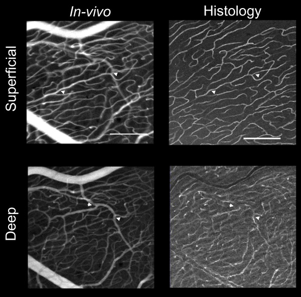
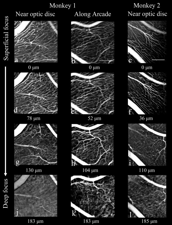
We injected sodium fluorescein intravenously in two macaque monkeys while imaging the retina with an FAO-SLO. An argon laser provided the 488 nm excitation source for fluorescence imaging. Reflectance images, obtained simultaneously with near infrared light, permitted precise surface registration of individual frames of the fluorescence imaging. In-vivo imaging was then compared to ex-vivo confocal microscopy of the same tissue.

RESULTS

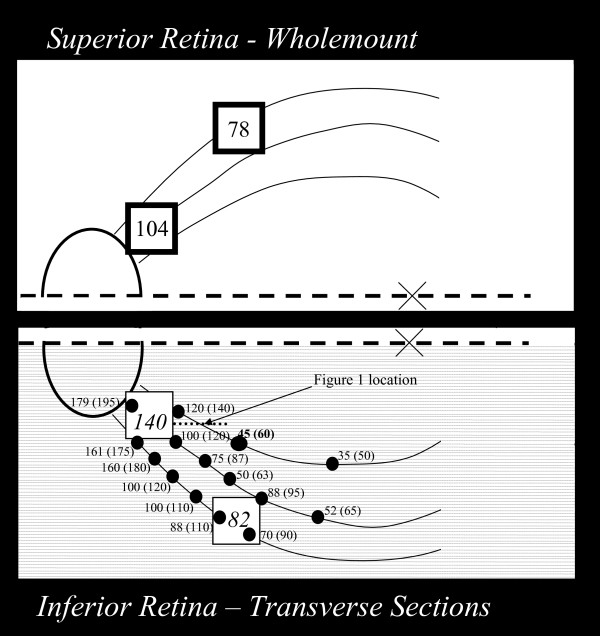
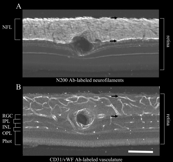
Superficial focus (innermost retina) at all depths within the NFL revealed a vasculature with extremely long capillaries, thin walls, little variation in caliber and parallel-linked structure oriented parallel to the NFL axons, typical of the radial peripapillary capillaries (RPCs). However, at a deeper focus beneath the NFL, (toward outer retina) the polygonal pattern typical of the ganglion cell layer (inner) and outer retinal vasculature was seen. These distinguishing patterns were also seen on histological examination of the same retinas. Furthermore, the thickness of the RPC beds and the caliber of individual RPCs determined by imaging closely matched that measured in histological sections.

CONCLUSION

This robust method demonstrates in-vivo, high-resolution, confocal imaging of the vasculature through the full thickness of the NFL in the living macaque, in precise agreement with histology. FAO provides a new tool to examine possible primary or secondary role of the nerve fiber layer vasculature in retinal vascular disorders and other eye diseases, such as glaucoma.

摘要

背景

尽管有人提出神经纤维层血管系统的改变可能参与包括青光眼在内的眼部疾病的病因,但一直无法在体内检查该血管系统。本报告描述了一种新型成像方法,即荧光自适应光学(FAO)扫描激光检眼镜(SLO),它首次实现了在活体猕猴体内对该血管系统进行成像,并比较了该血管床的体内和体外成像。

方法

我们对两只猕猴静脉注射荧光素钠,同时用FAO-SLO对视网膜进行成像。氩激光为荧光成像提供488nm激发源。与近红外光同时获得的反射图像允许对荧光成像的各个帧进行精确的表面配准。然后将体内成像与相同组织的体外共聚焦显微镜检查进行比较。

结果

在神经纤维层内所有深度的浅表焦点(最内层视网膜)显示出一个血管系统,其毛细血管极长、壁薄、管径变化小且平行连接结构与神经纤维层轴突平行,这是放射状视乳头周围毛细血管(RPC)的典型特征。然而,在神经纤维层下方更深的焦点处(朝向视网膜外层),可见神经节细胞层(内层)和视网膜外层血管系统典型的多边形模式。在相同视网膜的组织学检查中也观察到了这些独特模式。此外,通过成像确定的RPC床厚度和单个RPC的管径与组织学切片中测量的结果紧密匹配。

结论

这种可靠的方法在活体猕猴体内实现了对神经纤维层全厚度血管系统的高分辨率共聚焦成像,与组织学结果精确一致。FAO提供了一种新工具,可用于研究神经纤维层血管系统在视网膜血管疾病和其他眼部疾病(如青光眼)中可能的原发性或继发性作用。

https://cdn.ncbi.nlm.nih.gov/pmc/blobs/495d/2744910/4c7ddc41bd3e/1471-2415-9-9-4.jpg
https://cdn.ncbi.nlm.nih.gov/pmc/blobs/495d/2744910/c8b957998dd9/1471-2415-9-9-1.jpg
https://cdn.ncbi.nlm.nih.gov/pmc/blobs/495d/2744910/b90845aef1e0/1471-2415-9-9-2.jpg
https://cdn.ncbi.nlm.nih.gov/pmc/blobs/495d/2744910/80c2e7cec780/1471-2415-9-9-3.jpg
https://cdn.ncbi.nlm.nih.gov/pmc/blobs/495d/2744910/4c7ddc41bd3e/1471-2415-9-9-4.jpg

相似文献

1
In-vivo imaging of retinal nerve fiber layer vasculature: imaging histology comparison.视网膜神经纤维层脉管系统的体内成像:成像与组织学比较
BMC Ophthalmol. 2009 Aug 23;9:9. doi: 10.1186/1471-2415-9-9.
2
Label-Free Density Measurements of Radial Peripapillary Capillaries in the Human Retina.人视网膜中径向视乳头周围毛细血管的无标记密度测量
PLoS One. 2015 Aug 7;10(8):e0135151. doi: 10.1371/journal.pone.0135151. eCollection 2015.
3
In vivo imaging of microscopic structures in the rat retina.大鼠视网膜微观结构的体内成像。
Invest Ophthalmol Vis Sci. 2009 Dec;50(12):5872-9. doi: 10.1167/iovs.09-3675. Epub 2009 Jul 2.
4
Neural-vascular relationships in central retina of macaque monkeys (Macaca fascicularis).猕猴(食蟹猴)中央视网膜中的神经血管关系。
J Neurosci. 1992 Apr;12(4):1169-93. doi: 10.1523/JNEUROSCI.12-04-01169.1992.
5
In vivo imaging of the fine structure of rhodamine-labeled macaque retinal ganglion cells.若丹明标记的猕猴视网膜神经节细胞精细结构的体内成像。
Invest Ophthalmol Vis Sci. 2008 Jan;49(1):467-73. doi: 10.1167/iovs.07-0605.
6
Imaging individual neurons in the retinal ganglion cell layer of the living eye.对活体眼睛视网膜神经节细胞层中的单个神经元进行成像。
Proc Natl Acad Sci U S A. 2017 Jan 17;114(3):586-591. doi: 10.1073/pnas.1613445114. Epub 2017 Jan 3.
7
Morphology and topography of retinal pericytes in the living mouse retina using in vivo adaptive optics imaging and ex vivo characterization.利用活体自适应光学成像和离体特性分析研究活体小鼠视网膜内周细胞的形态和拓扑结构。
Invest Ophthalmol Vis Sci. 2013 Dec 19;54(13):8237-50. doi: 10.1167/iovs.13-12581.
8
In vivo optical imaging of human retinal capillary networks using speckle variance optical coherence tomography with quantitative clinico-histological correlation.使用散斑方差光学相干断层扫描对人视网膜毛细血管网络进行体内光学成像,并进行定量临床组织学相关性分析。
Microvasc Res. 2015 Jul;100:32-9. doi: 10.1016/j.mvr.2015.04.006. Epub 2015 Apr 24.
9
Quantitative Optical Coherence Tomography Angiography of Radial Peripapillary Capillaries in Glaucoma, Glaucoma Suspect, and Normal Eyes.青光眼、疑似青光眼和正常眼的视盘周围放射状毛细血管的定量光学相干断层扫描血管造影
Am J Ophthalmol. 2016 Oct;170:41-49. doi: 10.1016/j.ajo.2016.07.015. Epub 2016 Jul 25.
10
In vivo wide-field multispectral scanning laser ophthalmoscopy-optical coherence tomography mouse retinal imager: longitudinal imaging of ganglion cells, microglia, and Müller glia, and mapping of the mouse retinal and choroidal vasculature.体内宽视野多光谱扫描激光眼科检眼镜 - 光学相干断层扫描小鼠视网膜成像仪:神经节细胞、小胶质细胞和穆勒胶质细胞的纵向成像,以及小鼠视网膜和脉络膜血管系统的绘图
J Biomed Opt. 2015;20(12):126005. doi: 10.1117/1.JBO.20.12.126005.

引用本文的文献

1
Quantitative analysis of peripapillary RNFL capillary density and thickness in patients with mild nonproliferative diabetic retinopathy.轻度非增殖性糖尿病视网膜病变患者视乳头周围视网膜神经纤维层毛细血管密度和厚度的定量分析。
Front Endocrinol (Lausanne). 2025 Jan 24;15:1404157. doi: 10.3389/fendo.2024.1404157. eCollection 2024.
2
Vascular density normative data of radial peripapillary capillary plexus in healthy Caucasian subjects.健康高加索受试者视乳头周围视网膜毛细血管丛的血管密度标准数据。
Graefes Arch Clin Exp Ophthalmol. 2025 Jan;263(1):97-103. doi: 10.1007/s00417-024-06623-6. Epub 2024 Aug 30.
3
Quantifying peripapillary vessel density and retinal nerve fibre layer in type 1 diabetic children without clinically detectable retinopathy using OCTA.

本文引用的文献

1
In vivo fluorescence imaging of primate retinal ganglion cells and retinal pigment epithelial cells.灵长类动物视网膜神经节细胞和视网膜色素上皮细胞的体内荧光成像。
Opt Express. 2006 Aug 7;14(16):7144-58. doi: 10.1364/oe.14.007144.
2
Retinal peripapillary nerve fiber layer thickness in neuromyelitis optica.视神经脊髓炎中视网膜视乳头周围神经纤维层厚度
Invest Ophthalmol Vis Sci. 2008 Oct;49(10):4412-7. doi: 10.1167/iovs.08-1815. Epub 2008 Jul 9.
3
Relative course of retinal nerve fiber layer birefringence and thickness and retinal function changes after optic nerve transection.
使用光学相干断层扫描血管造影术(OCTA)对无临床可检测视网膜病变的1型糖尿病儿童的视乳头周围血管密度和视网膜神经纤维层进行量化。
Int J Ophthalmol. 2024 Feb 18;17(2):278-281. doi: 10.18240/ijo.2024.02.08. eCollection 2024.
4
Changes of Peripapillary Capillary Density in Patients with Vogt-Koyanagi-Harada Disease Evaluated by Optical Coherence Tomography Angiography.通过光学相干断层扫描血管造影术评估的Vogt-小柳-原田病患者视乳头周围毛细血管密度的变化
J Ophthalmol. 2023 Apr 17;2023:1271070. doi: 10.1155/2023/1271070. eCollection 2023.
5
Comparison of peripapillary capillary plexus using optical coherence tomography angiography and retinal nerve fibre layer analysis using spectral domain optical coherence tomography in glaucoma patients, glaucoma suspects, and healthy subjects.青光眼患者、疑似青光眼患者和健康受试者的光学相干断层扫描血管造影的视盘周围毛细血管丛与光谱域光学相干断层扫描的视网膜神经纤维层分析比较。
Indian J Ophthalmol. 2022 Dec;70(12):4146-4151. doi: 10.4103/ijo.IJO_1456_22.
6
Reduced choroidal peripapillary capillaries in thyroid-associated ophthalmopathy with early stage of dysthyroid optic neuropathy.甲状腺相关性眼病伴甲状腺功能异常性视神经病变早期脉络膜视乳头周围毛细血管减少
Int J Ophthalmol. 2022 Jul 18;15(7):1135-1141. doi: 10.18240/ijo.2022.07.14. eCollection 2022.
7
Adaptive optics scanning laser ophthalmoscopy may support early diagnosis of glaucoma.自适应光学扫描激光检眼镜可辅助青光眼的早期诊断。
Indian J Ophthalmol. 2022 Aug;70(8):2877-2882. doi: 10.4103/ijo.IJO_1044_21.
8
Evaluation of Radial Peripapillary Capillary Density in G6PD Deficiency: An OCT Angiography Pilot Study.葡萄糖-6-磷酸脱氢酶缺乏症患者的视盘周围视网膜毛细血管密度评估:一项光学相干断层扫描血管造影初步研究
J Clin Med. 2022 Jun 8;11(12):3282. doi: 10.3390/jcm11123282.
9
Longitudinal In Vivo Changes in Radial Peripapillary Capillaries and Optic Nerve Head Structure in Non-Human Primates With Early Experimental Glaucoma.早期实验性青光眼非人类灵长类动物视乳头周围毛细血管和视神经头部结构的纵向活体变化。
Invest Ophthalmol Vis Sci. 2022 Jan 3;63(1):10. doi: 10.1167/iovs.63.1.10.
10
Cell - Vessel Mismatch in Glaucoma: Correlation of Ganglion Cell Layer Soma and Capillary Densities.青光眼的细胞-血管不匹配:神经节细胞层体和毛细血管密度的相关性。
Invest Ophthalmol Vis Sci. 2021 Oct 4;62(13):2. doi: 10.1167/iovs.62.13.2.
视神经横断后视网膜神经纤维层双折射和厚度的相对变化过程以及视网膜功能改变
Invest Ophthalmol Vis Sci. 2008 Oct;49(10):4444-52. doi: 10.1167/iovs.08-2255. Epub 2008 Jun 19.
4
Light-induced retinal changes observed with high-resolution autofluorescence imaging of the retinal pigment epithelium.通过视网膜色素上皮的高分辨率自发荧光成像观察到的光诱导视网膜变化。
Invest Ophthalmol Vis Sci. 2008 Aug;49(8):3715-29. doi: 10.1167/iovs.07-1430. Epub 2008 Apr 11.
5
In vivo imaging of the fine structure of rhodamine-labeled macaque retinal ganglion cells.若丹明标记的猕猴视网膜神经节细胞精细结构的体内成像。
Invest Ophthalmol Vis Sci. 2008 Jan;49(1):467-73. doi: 10.1167/iovs.07-0605.
6
A framework for comparing structural and functional measures of glaucomatous damage.一种用于比较青光眼性损害的结构和功能测量指标的框架。
Prog Retin Eye Res. 2007 Nov;26(6):688-710. doi: 10.1016/j.preteyeres.2007.08.001. Epub 2007 Aug 21.
7
Glaucoma: macrocosm to microcosm the Friedenwald lecture.青光眼:从宏观到微观——弗里登瓦尔德讲座
Invest Ophthalmol Vis Sci. 2005 Aug;46(8):2662-70. doi: 10.1167/iovs.04-1070.
8
In vivo dynamics of retinal injury and repair in the rhodopsin mutant dog model of human retinitis pigmentosa.人类色素性视网膜炎视紫红质突变犬模型中视网膜损伤与修复的体内动态变化
Proc Natl Acad Sci U S A. 2005 Apr 5;102(14):5233-8. doi: 10.1073/pnas.0408892102. Epub 2005 Mar 22.
9
Histological measurement of retinal nerve fibre layer thickness.视网膜神经纤维层厚度的组织学测量。
Eye (Lond). 2005 May;19(5):491-8. doi: 10.1038/sj.eye.6701569.
10
Detecting early glaucoma by assessment of retinal nerve fiber layer thickness and visual function.通过评估视网膜神经纤维层厚度和视觉功能来检测早期青光眼。
Invest Ophthalmol Vis Sci. 2001 Aug;42(9):1993-2003.