V S T Glaucoma Center, Dr. Kallam Anji Reddy Campus, L. V. Prasad Eye Institute, Hyderabad, Telangana, India.
Engineering Group, Dr. Kallam Anji Reddy campus, L. V. Prasad Eye Institute, Hyderabad, Telangana, India.
Indian J Ophthalmol. 2022 Aug;70(8):2877-2882. doi: 10.4103/ijo.IJO_1044_21.
To compare image characteristics of retinal nerve fiber layer (RNFL) between glaucoma patients and healthy controls using adaptive optics scanning laser ophthalmoscopy (AOSLO).
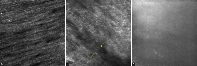
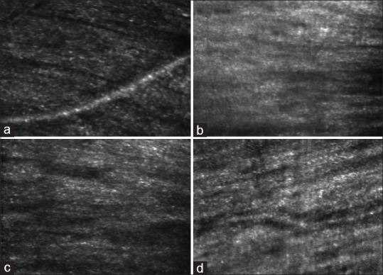
This was a cross-sectional pilot study with two groups: a glaucoma group with patients with moderate or severe glaucoma as per the Hodapp-Parrish-Anderson classification system and a control group with healthy individuals. The optic nerve damage in moderate glaucoma was predominantly located in only one hemisphere; the other hemisphere was un- or minimally affected on optical coherence tomography and automated perimetry and is referred to as early glaucoma. The structure of RNFL bundles and gain (%) in RNFL images with mean pixel values between 15 and 35 were analyzed. Imaging was performed one degree away from the optic disc margin at two and four cardinal clock positions in the glaucoma and control groups, respectively. The field of view was 1.3° at 2.3 μ resolution. We studied one eye per participant.
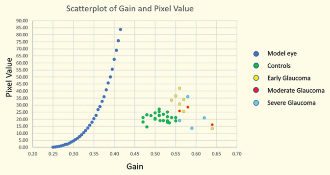
There were 11 glaucoma patients and 7 healthy controls. Imaging was successful at 88% of the locations in controls and early glaucoma; the reflectivity differed significantly (0.51 and 0.56, respectively, P < 0.001) but not the structure of RNFL bundles (Cohen's Kappa 0.11) between them. In patients with moderate and severe glaucoma, imaging was successful only at 46% of the locations; RNFL bundles were not discernible, and RNFL reflectivity did not differ from those with early glaucoma (P < 0.11).
The recorded gain (%) of RNFL images obtained using AOSLO could be an objective indicator of early glaucoma.
使用自适应光学扫描激光检眼镜(AOSLO)比较青光眼患者和健康对照者的视网膜神经纤维层(RNFL)图像特征。
这是一项横断面试点研究,分为两组:一组为青光眼组,患者为根据 Hodapp-Parrish-Anderson 分类系统诊断的中重度青光眼;另一组为对照组,患者为健康个体。中重度青光眼的视神经损伤主要局限于一个半球;另一个半球在光学相干断层扫描和自动视野计上未受影响或仅轻度受累,称为早期青光眼。分析平均像素值在 15 到 35 之间的 RNFL 束结构和 RNFL 图像增益(%)。在青光眼组和对照组,分别在距视盘边缘 1 度处和两个及四个钟点位置进行成像。视野分辨率为 2.3 μ,为 1.3°。每位参与者研究一只眼。
共有 11 名青光眼患者和 7 名健康对照者。在对照组和早期青光眼患者中,有 88%的位置可以成功成像;反射率差异有统计学意义(分别为 0.51 和 0.56,P<0.001),但 RNFL 束结构无差异(Cohen's Kappa 0.11)。在中重度青光眼患者中,只有 46%的位置可以成功成像;RNFL 束不可见,RNFL 反射率与早期青光眼患者无差异(P<0.11)。
AOSLO 获得的 RNFL 图像记录的增益(%)可能是早期青光眼的客观指标。