• 文献检索
  • 文档翻译
  • 深度研究
  • 学术资讯
  • Suppr Zotero 插件Zotero 插件
  • 邀请有礼
  • 套餐&价格
  • 历史记录
应用&插件
Suppr Zotero 插件Zotero 插件浏览器插件Mac 客户端Windows 客户端微信小程序
定价
高级版会员购买积分包购买API积分包
服务
文献检索文档翻译深度研究API 文档MCP 服务
关于我们
关于 Suppr公司介绍联系我们用户协议隐私条款
关注我们

Suppr 超能文献

核心技术专利:CN118964589B侵权必究
粤ICP备2023148730 号-1Suppr @ 2026

文献检索

告别复杂PubMed语法,用中文像聊天一样搜索,搜遍4000万医学文献。AI智能推荐,让科研检索更轻松。

立即免费搜索

文件翻译

保留排版,准确专业,支持PDF/Word/PPT等文件格式,支持 12+语言互译。

免费翻译文档

深度研究

AI帮你快速写综述,25分钟生成高质量综述,智能提取关键信息,辅助科研写作。

立即免费体验

使用 MDCT 评估小梁骨结构:以高分辨率 pQCT 作为参考标准比较 64 层和 320 层 CT。

Assessment of trabecular bone structure using MDCT: comparison of 64- and 320-slice CT using HR-pQCT as the reference standard.

机构信息

Department of Radiology, Charité Campus Mitte, Universitaetsmedizin, Berlin, Germany.

出版信息

Eur Radiol. 2010 Feb;20(2):458-68. doi: 10.1007/s00330-009-1571-7. Epub 2009 Aug 27.

DOI:10.1007/s00330-009-1571-7
PMID:19711081
原文链接:https://pmc.ncbi.nlm.nih.gov/articles/PMC2814042/
Abstract

OBJECTIVES

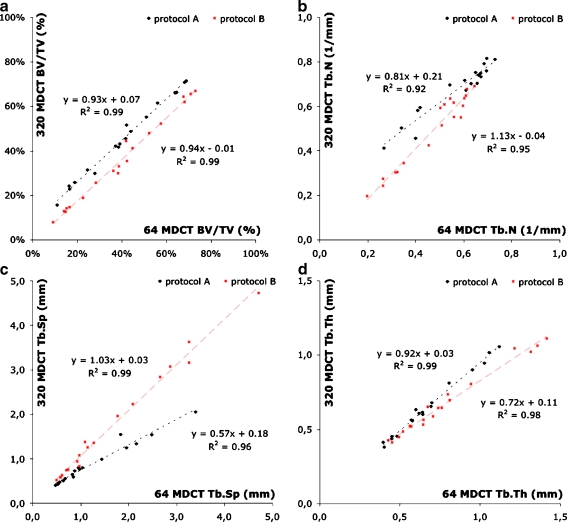
The aim of our study was to perform trabecular bone structure analysis with images from 64- and 320-slice multidetector computed tomography (MDCT) and to compare these with high-resolution peripheral computed tomography (HR-pQCT).

MATERIALS AND METHODS

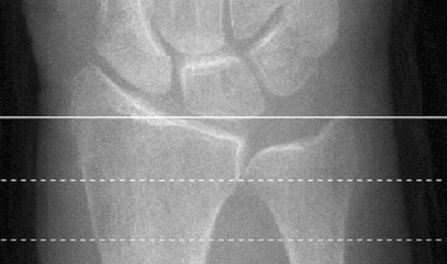
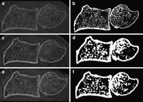
Twenty human cadaver distal forearm specimens were imaged on a 64- and 320-slice MDCT system at 120 kVp, 200 mA and 135 kVp, 400 mA (in-plane pixel size 234 microm; slice thickness 500 microm). HR-pQCT imaging was performed at an isotropic voxel size of 41 microm. Bone volume fraction (BV/TV), trabecular number (Tb.N), thickness (Tb.Th) and separation (Tb.Sp) were computed.

RESULTS

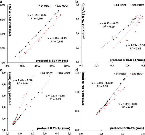
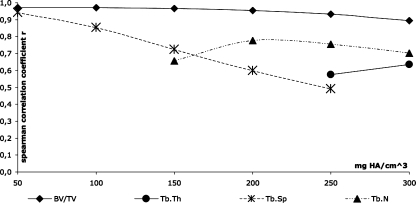
MDCT-derived BV/TV and Tb.Sp were highly correlated (r = 0.92-0.96, p < 0.0001) with the corresponding HR-pQCT parameters. Tb.Th was the only structure measure that did not yield any significant correlation.

CONCLUSION

The 64- and 320-slice MDCT systems both perform equally well in depicting trabecular bone architecture. However, because of constrained resolutions accurate derivation of trabecular bone measures is limited to only a subset of microarchitectural parameters.

摘要

目的

我们的研究旨在使用 64 层和 320 层多层螺旋 CT(MDCT)的图像进行小梁骨结构分析,并将其与高分辨率外周 CT(HR-pQCT)进行比较。

材料与方法

20 个人体尸检的远侧前臂标本在 120kVp、200mA 和 135kVp、400mA 的 64 层和 320 层 MDCT 系统上进行成像(平面像素大小 234μm;层厚 500μm)。HR-pQCT 成像的各向同性体素大小为 41μm。计算骨体积分数(BV/TV)、小梁数(Tb.N)、厚度(Tb.Th)和分离度(Tb.Sp)。

结果

MDCT 衍生的 BV/TV 和 Tb.Sp 与相应的 HR-pQCT 参数高度相关(r=0.92-0.96,p<0.0001)。Tb.Th 是唯一没有显著相关性的结构测量指标。

结论

64 层和 320 层 MDCT 系统在描绘小梁骨结构方面表现相当。然而,由于分辨率受限,小梁骨测量的准确推导仅限于微观结构参数的一个子集。

https://cdn.ncbi.nlm.nih.gov/pmc/blobs/c45f/2814042/a8586b197b22/330_2009_1571_Fig5_HTML.jpg
https://cdn.ncbi.nlm.nih.gov/pmc/blobs/c45f/2814042/cbb5e3298e10/330_2009_1571_Fig1_HTML.jpg
https://cdn.ncbi.nlm.nih.gov/pmc/blobs/c45f/2814042/611bab23fe80/330_2009_1571_Fig2_HTML.jpg
https://cdn.ncbi.nlm.nih.gov/pmc/blobs/c45f/2814042/4a54cad1050a/330_2009_1571_Fig3_HTML.jpg
https://cdn.ncbi.nlm.nih.gov/pmc/blobs/c45f/2814042/2c8d652e28d9/330_2009_1571_Fig4_HTML.jpg
https://cdn.ncbi.nlm.nih.gov/pmc/blobs/c45f/2814042/a8586b197b22/330_2009_1571_Fig5_HTML.jpg

相似文献

1
Assessment of trabecular bone structure using MDCT: comparison of 64- and 320-slice CT using HR-pQCT as the reference standard.使用 MDCT 评估小梁骨结构:以高分辨率 pQCT 作为参考标准比较 64 层和 320 层 CT。
Eur Radiol. 2010 Feb;20(2):458-68. doi: 10.1007/s00330-009-1571-7. Epub 2009 Aug 27.
2
Human trabecular bone microarchitecture can be assessed independently of density with second generation HR-pQCT.利用第二代高分辨率外周定量计算机断层扫描(HR-pQCT),可以独立于骨密度评估人类小梁骨微结构。
Bone. 2015 Oct;79:213-21. doi: 10.1016/j.bone.2015.06.006. Epub 2015 Jun 14.
3
Validation of calcaneus trabecular microstructure measurements by HR-pQCT.跟骨小梁微结构测量的 HR-pQCT 验证。
Bone. 2018 Jan;106:69-77. doi: 10.1016/j.bone.2017.09.013. Epub 2017 Oct 3.
4
The effect of voxel size on high-resolution peripheral computed tomography measurements of trabecular and cortical bone microstructure.体素大小对高分辨率外周计算机断层扫描测量骨小梁和皮质骨微观结构的影响。
Med Phys. 2012 Apr;39(4):1893-903. doi: 10.1118/1.3689813.
5
High-resolution peripheral quantitative computed tomography can assess microstructural and mechanical properties of human distal tibial bone.高分辨率外周定量计算机断层扫描可评估人类胫骨远端骨的微观结构和力学性能。
J Bone Miner Res. 2010 Apr;25(4):746-56. doi: 10.1359/jbmr.090822.
6
Trabecular bone structure analysis in the osteoporotic spine using a clinical in vivo setup for 64-slice MDCT imaging: comparison to microCT imaging and microFE modeling.使用用于64层MDCT成像的临床体内装置对骨质疏松性脊柱小梁骨结构进行分析:与显微CT成像和微观有限元建模的比较。
J Bone Miner Res. 2009 Sep;24(9):1628-37. doi: 10.1359/jbmr.090311.
7
In vivo determination of bone structure in postmenopausal women: a comparison of HR-pQCT and high-field MR imaging.绝经后女性骨结构的体内测定:高分辨率外周定量计算机断层扫描与高场磁共振成像的比较
J Bone Miner Res. 2008 Apr;23(4):463-74. doi: 10.1359/jbmr.071116.
8
Comparison of pQCT-based measures of radial bone geometry and apparent trabecular bone structure using manufacturer and in-house-developed algorithms.基于 pQCT 的桡骨骨几何和表观骨小梁结构测量值的比较,使用制造商和内部开发的算法。
J Clin Densitom. 2010 Oct-Dec;13(4):433-40. doi: 10.1016/j.jocd.2010.06.007. Epub 2010 Sep 17.
9
Quantitative imaging of peripheral trabecular bone microarchitecture using MDCT.使用多层螺旋CT对外周小梁骨微结构进行定量成像。
Med Phys. 2018 Jan;45(1):236-249. doi: 10.1002/mp.12632. Epub 2017 Nov 23.
10
Comparison of synchrotron radiation and conventional x-ray microcomputed tomography for assessing trabecular bone microarchitecture of human femoral heads.同步辐射与传统X射线显微计算机断层扫描在评估人股骨头小梁骨微结构方面的比较。
Med Phys. 2006 Sep;33(9):3568-77. doi: 10.1118/1.2256069.

引用本文的文献

1
Cone beam computed tomography vs. microcomputed tomography for trabecular bone healing assessment : An ex vivo study in rabbit mandibles.锥形束计算机断层扫描与微型计算机断层扫描用于小梁骨愈合评估的比较:兔下颌骨的体外研究
J Orofac Orthop. 2025 May 20. doi: 10.1007/s00056-025-00589-8.
2
Evaluation of trabecular bone microstructure and cortical morphology using cone-beam and micro-CT images: impact of tube voltage setting.使用锥束CT和显微CT图像评估小梁骨微观结构和皮质形态:管电压设置的影响
BMC Oral Health. 2025 Apr 29;25(1):664. doi: 10.1186/s12903-025-05855-0.
3
Bone remodeling simulation using spatial influence function in macroscopic cube case.

本文引用的文献

1
Feasibility of measuring trabecular bone structure of the proximal femur using 64-slice multidetector computed tomography in a clinical setting.在临床环境中使用64层多层螺旋计算机断层扫描测量股骨近端小梁骨结构的可行性。
Calcif Tissue Int. 2008 Nov;83(5):332-41. doi: 10.1007/s00223-008-9181-y. Epub 2008 Oct 15.
2
In vivo trabecular bone morphologic and mechanical relationship using high-resolution 3-T MRI.使用高分辨率3-T磁共振成像研究体内小梁骨形态与力学的关系。
AJR Am J Roentgenol. 2008 Sep;191(3):721-6. doi: 10.2214/AJR.07.3528.
3
High-resolution pQCT analysis at the distal radius and tibia discriminates patients with recent wrist and femoral neck fractures.
在宏观立方体案例中使用空间影响函数的骨重塑模拟。
Front Bioeng Biotechnol. 2024 Nov 29;12:1498812. doi: 10.3389/fbioe.2024.1498812. eCollection 2024.
4
Photon-counting detector CT and energy-integrating detector CT for trabecular bone microstructure analysis of cubic specimens from human radius.光子计数探测器 CT 和能量积分探测器 CT 用于分析人桡骨立方样本的小梁骨微结构。
Eur Radiol Exp. 2022 Jul 27;6(1):31. doi: 10.1186/s41747-022-00286-w.
5
Effect of different voxel sizes on the accuracy of CBCT measurements of trabecular bone microstructure: A comparative micro-CT study.不同体素大小对锥形束计算机断层扫描测量小梁骨微结构准确性的影响:一项对比性显微计算机断层扫描研究。
Imaging Sci Dent. 2022 Jun;52(2):171-179. doi: 10.5624/isd.20220025. Epub 2022 Apr 28.
6
Dose-efficient assessment of trabecular microstructure using ultra-high-resolution photon-counting CT.利用超高分辨率光子计数 CT 进行高效的小梁微结构评估。
Z Med Phys. 2022 Nov;32(4):403-416. doi: 10.1016/j.zemedi.2022.04.001. Epub 2022 May 18.
7
In-Vivo Degradation Behavior and Osseointegration of 3D Powder-Printed Calcium Magnesium Phosphate Cement Scaffolds.3D粉末打印磷酸钙镁骨水泥支架的体内降解行为及骨整合
Materials (Basel). 2021 Feb 17;14(4):946. doi: 10.3390/ma14040946.
8
A comparative study of trabecular bone micro-structural measurements using different CT modalities.不同 CT 模态下小梁骨微结构测量的对比研究。
Phys Med Biol. 2020 Nov 27;65(23). doi: 10.1088/1361-6560/abc367.
9
Vertebral body insufficiency fractures: detection of vertebrae at risk on standard CT images using texture analysis and machine learning.椎体骨量不足性骨折:使用纹理分析和机器学习技术在标准 CT 图像上检测有风险的椎体。
Eur Radiol. 2019 May;29(5):2207-2217. doi: 10.1007/s00330-018-5846-8. Epub 2018 Dec 5.
10
Penalized-Likelihood Reconstruction With High-Fidelity Measurement Models for High-Resolution Cone-Beam Imaging.基于高保真测量模型的 penalized-likelihood 重建技术在高分率锥形束成像中的应用。
IEEE Trans Med Imaging. 2018 Apr;37(4):988-999. doi: 10.1109/TMI.2017.2779406.
桡骨远端和胫骨的高分辨率外周定量计算机断层扫描分析可鉴别近期发生手腕和股骨颈骨折的患者。
J Bone Miner Res. 2008 Nov;23(11):1741-50. doi: 10.1359/jbmr.080704.
4
Adaptations in trabecular bone microarchitecture in Olympic athletes determined by 7T MRI.通过7T磁共振成像确定的奥运会运动员小梁骨微结构的适应性变化。
J Magn Reson Imaging. 2008 May;27(5):1089-95. doi: 10.1002/jmri.21326.
5
In vivo ultra-high-field magnetic resonance imaging of trabecular bone microarchitecture at 7 T.7T下小梁骨微结构的体内超高场磁共振成像
J Magn Reson Imaging. 2008 Apr;27(4):854-9. doi: 10.1002/jmri.21325.
6
Mechanisms of the anabolic effects of teriparatide on bone: insight from the treatment of a patient with pycnodysostosis.特立帕肽对骨骼合成代谢作用的机制:来自一名致密性成骨不全症患者治疗的见解
J Bone Miner Res. 2008 Jul;23(7):1076-83. doi: 10.1359/jbmr.080231.
7
Resolution dependence of the non-metric trabecular structure indices.非度量小梁结构指数的分辨率依赖性
Bone. 2008 Apr;42(4):728-36. doi: 10.1016/j.bone.2007.12.004. Epub 2007 Dec 23.
8
Wrist fracture as a predictor of future fractures in younger versus older postmenopausal women: results from the National Osteoporosis Risk Assessment (NORA).手腕骨折作为绝经后年轻女性与老年女性未来骨折的预测指标:来自国家骨质疏松症风险评估(NORA)的结果。
Osteoporos Int. 2008 May;19(5):607-13. doi: 10.1007/s00198-007-0508-8. Epub 2007 Dec 6.
9
In vivo determination of bone structure in postmenopausal women: a comparison of HR-pQCT and high-field MR imaging.绝经后女性骨结构的体内测定:高分辨率外周定量计算机断层扫描与高场磁共振成像的比较
J Bone Miner Res. 2008 Apr;23(4):463-74. doi: 10.1359/jbmr.071116.
10
Assessment of trabecular bone structure comparing magnetic resonance imaging at 3 Tesla with high-resolution peripheral quantitative computed tomography ex vivo and in vivo.比较3特斯拉磁共振成像与高分辨率外周定量计算机断层扫描在体外和体内对小梁骨结构的评估。
Osteoporos Int. 2008 May;19(5):653-61. doi: 10.1007/s00198-007-0495-9. Epub 2007 Nov 9.