Mienaltowski Michael J, Huang Liping, Frisbie David D, McIlwraith C Wayne, Stromberg Arnold J, Bathke Arne C, Macleod James N
University of Kentucky, Department of Veterinary Science, Maxwell H, Gluck Equine Research Center, Lexington, KY 40546-0099, USA.
BMC Med Genomics. 2009 Sep 14;2:60. doi: 10.1186/1755-8794-2-60.
Full-thickness articular cartilage lesions that reach to the subchondral bone yet are restricted to the chondral compartment usually fill with a fibrocartilage-like repair tissue which is structurally and biomechanically compromised relative to normal articular cartilage. The objective of this study was to evaluate transcriptional differences between chondrocytes of normal articular cartilage and repair tissue cells four months post-microfracture.
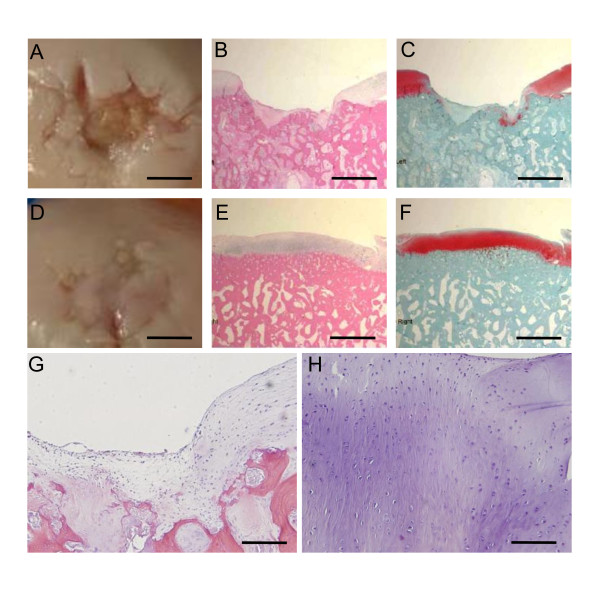
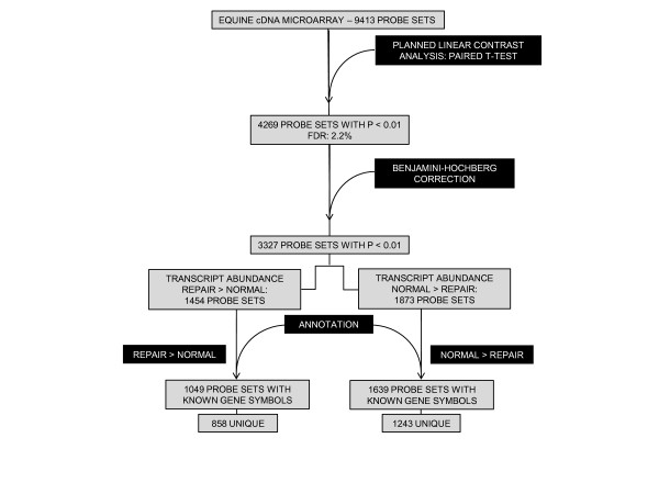
Bilateral one-cm2 full-thickness defects were made in the articular surface of both distal femurs of four adult horses followed by subchondral microfracture. Four months postoperatively, repair tissue from the lesion site and grossly normal articular cartilage from within the same femorotibial joint were collected. Total RNA was isolated from the tissue samples, linearly amplified, and applied to a 9,413-probe set equine-specific cDNA microarray. Eight paired comparisons matched by limb and horse were made with a dye-swap experimental design with validation by histological analyses and quantitative real-time polymerase chain reaction (RT-qPCR).
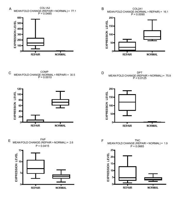
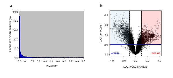
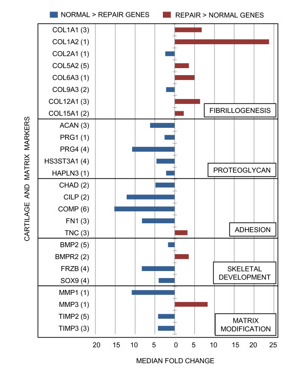
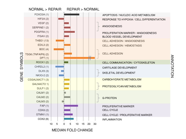
Statistical analyses revealed 3,327 (35.3%) differentially expressed probe sets. Expression of biomarkers typically associated with normal articular cartilage and fibrocartilage repair tissue corroborate earlier studies. Other changes in gene expression previously unassociated with cartilage repair were also revealed and validated by RT-qPCR.
The magnitude of divergence in transcriptional profiles between normal chondrocytes and the cells that populate repair tissue reveal substantial functional differences between these two cell populations. At the four-month postoperative time point, the relative deficiency within repair tissue of gene transcripts which typically define articular cartilage indicate that while cells occupying the lesion might be of mesenchymal origin, they have not recapitulated differentiation to the chondrogenic phenotype of normal articular chondrocytes.
全层关节软骨损伤累及软骨下骨,但局限于软骨层,通常会填充一种纤维软骨样修复组织,该组织在结构和生物力学方面相对于正常关节软骨存在缺陷。本研究的目的是评估正常关节软骨细胞与微骨折后四个月修复组织细胞之间的转录差异。
在四匹成年马双侧股骨远端关节表面制作直径1厘米的全层缺损,随后进行软骨下微骨折。术后四个月,收集损伤部位的修复组织以及同一股胫关节内大体正常的关节软骨。从组织样本中提取总RNA,进行线性扩增,并应用于一个包含9413个探针组的马特异性cDNA微阵列。采用染料交换实验设计,对八对按肢体和马匹匹配的样本进行比较,并通过组织学分析和定量实时聚合酶链反应(RT-qPCR)进行验证。
统计分析显示有3327个(35.3%)探针组表达存在差异。与正常关节软骨和纤维软骨修复组织相关的生物标志物表达情况证实了早期研究结果。RT-qPCR还揭示并验证了其他先前与软骨修复无关的基因表达变化。
正常软骨细胞与填充修复组织的细胞之间转录谱的差异程度表明这两个细胞群体之间存在显著的功能差异。在术后四个月的时间点,修复组织中通常定义关节软骨的基因转录本相对缺乏,这表明占据损伤部位的细胞可能起源于间充质,但它们尚未重现向正常关节软骨细胞软骨生成表型的分化。