Volbrecht Vicki J, Shrago Erin E, Schefrin Brooke E, Werner John S
Colorado State University, Ft. Collins, Colorado.
Color Res Appl. 2000 Dec 27;26(51):S32-S35. doi: 10.1002/1520-6378(2001)26:1+<::AID-COL8>3.0.CO;2-V.
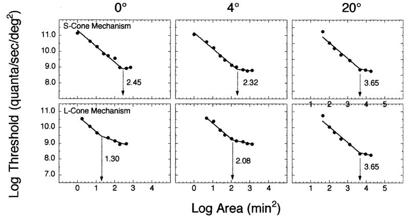
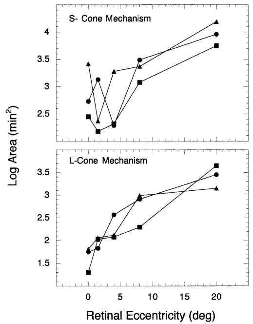
The purposes of this study were to measure areas of complete spatial summation (i.e., Ricco's area) for S- and L-cone mechanisms and to evaluate whether the sizes of Ricco's area could be explained in terms of either the densities of photoreceptors or ganglion cells. Increment thresholds were measured at the fovea and at 1.5°, 4°, 8°, and 20° in the superior retina using a temporal two-alternative forced-choice procedure. Test stimuli ranging from -0.36 to 4.61 log area (min(2)) were presented on concentric 12.3° adapting and auxiliary fields, which isolated either an S- or L-cone mechanism on the plateau of the respective threshold vs. intensity function. The data indicate that from 0-20° retinal eccentricity, the size of Ricco's area is larger for the S-cone mechanism than the L-cone mechanism, increases monotonically for the L-cone mechanism, and, for both cone mechanisms, increases between 8-20° retinal eccentricity. This latter finding suggests that ganglion cell density rather than cone density defines the size of Ricco's area in the parafoveal and peripheral retina.
本研究的目的是测量S-视锥细胞和L-视锥细胞机制的完全空间总和区域(即里科区域),并评估里科区域的大小是否可以用光感受器或神经节细胞的密度来解释。使用时间二择一强制选择程序,在中央凹以及视网膜上方1.5°、4°、8°和20°处测量增量阈值。在同心的12.3°适应场和辅助场中呈现范围从-0.36至4.61对数面积(min(2))的测试刺激,这些场在各自的阈值与强度函数的平稳段分离出S-视锥细胞或L-视锥细胞机制。数据表明,在视网膜偏心度0 - 20°范围内,S-视锥细胞机制的里科区域大小比L-视锥细胞机制的大,L-视锥细胞机制的里科区域大小单调增加,并且对于两种视锥细胞机制,在视网膜偏心度8 - 20°之间里科区域大小增加。后一发现表明,在中央凹旁和周边视网膜中,神经节细胞密度而非视锥细胞密度决定了里科区域的大小。