• 文献检索
  • 文档翻译
  • 深度研究
  • 学术资讯
  • Suppr Zotero 插件Zotero 插件
  • 邀请有礼
  • 套餐&价格
  • 历史记录
应用&插件
Suppr Zotero 插件Zotero 插件浏览器插件Mac 客户端Windows 客户端微信小程序
定价
高级版会员购买积分包购买API积分包
服务
文献检索文档翻译深度研究API 文档MCP 服务
关于我们
关于 Suppr公司介绍联系我们用户协议隐私条款
关注我们

Suppr 超能文献

核心技术专利:CN118964589B侵权必究
粤ICP备2023148730 号-1Suppr @ 2026

文献检索

告别复杂PubMed语法,用中文像聊天一样搜索,搜遍4000万医学文献。AI智能推荐,让科研检索更轻松。

立即免费搜索

文件翻译

保留排版,准确专业,支持PDF/Word/PPT等文件格式,支持 12+语言互译。

免费翻译文档

深度研究

AI帮你快速写综述,25分钟生成高质量综述,智能提取关键信息,辅助科研写作。

立即免费体验

首次冠状动脉闭塞患者梗死演变的研究:与不同物种的比较——对心肌挽救评估的意义

Infarct evolution in man studied in patients with first-time coronary occlusion in comparison to different species - implications for assessment of myocardial salvage.

作者信息

Hedström Erik, Engblom Henrik, Frogner Fredrik, Aström-Olsson Karin, Ohlin Hans, Jovinge Stefan, Arheden Håkan

机构信息

Department of Clinical Physiology, Lund University Hospital, SE-221 85 Lund, Sweden.

出版信息

J Cardiovasc Magn Reson. 2009 Sep 23;11(1):38. doi: 10.1186/1532-429X-11-38.

DOI:10.1186/1532-429X-11-38
PMID:19775428
原文链接:https://pmc.ncbi.nlm.nih.gov/articles/PMC2758877/
Abstract

BACKGROUND

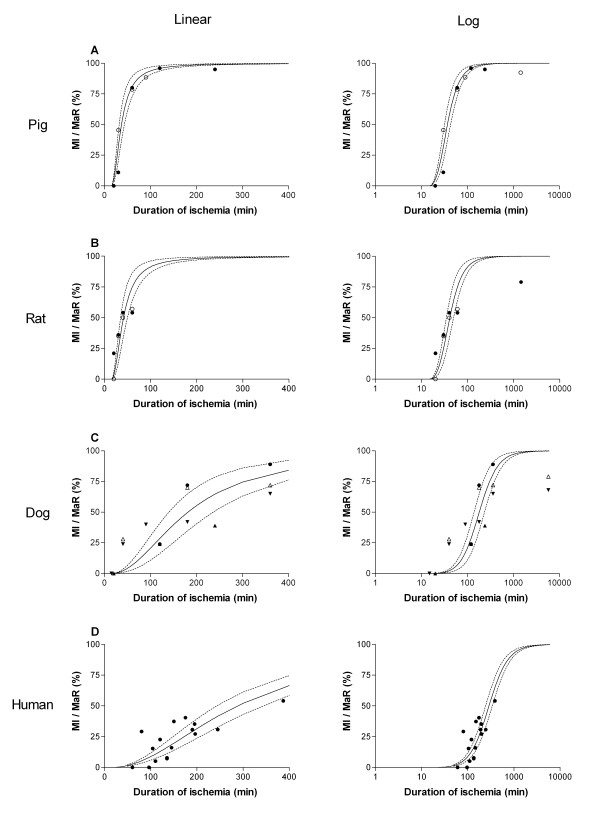
The time course of infarct evolution, i.e. how fast myocardial infarction (MI) develops during coronary artery occlusion, is well known for several species, whereas no direct evidence exists on the evolution of MI size normalized to myocardium at risk (MaR) in man. Despite the lack of direct evidence, current literature often refers to the "golden hour" as the time during which myocardial salvage can be accomplished by reperfusion therapy. Therefore, the aim of the present study was to investigate how duration of myocardial ischemia affects infarct evolution in man in relation to previous animal data. Consecutive patients with clinical signs of acute myocardial ischemia were screened and considered for enrollment. Particular care was taken to assure uniformity of the patients enrolled with regard to old MI, success of revascularization, collateral flow, release of biochemical markers prior to intervention etc. Sixteen patients were ultimately included in the study. Myocardium at risk was assessed acutely by acute myocardial perfusion single photon emission computed tomography (MPS) and by T2 imaging (T2-STIR) cardiovascular magnetic resonance (CMR) after one week in 10 of the 16 patients. Infarct size was measured by late gadolinium enhancement (LGE) at one week.

RESULTS

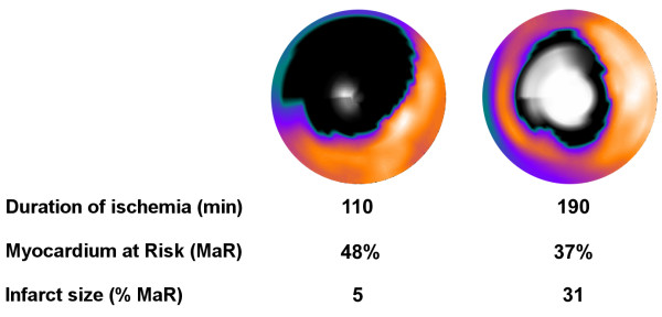
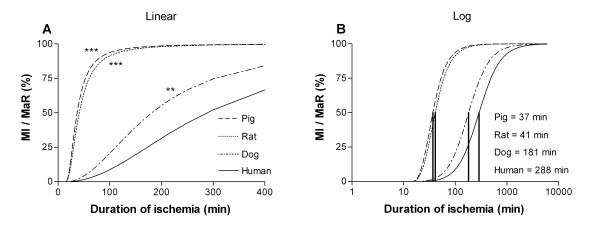
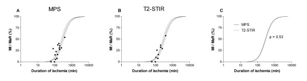
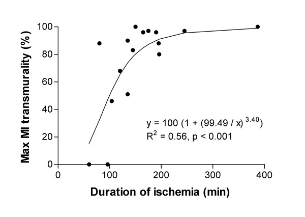
The time to reach 50% MI of the MaR (T50) was significantly shorter in pigs (37 min), rats (41 min) and dogs (181 min) compared to humans (288 min). There was no significant difference in T50 when using MPS compared to T2-STIR (p = 0.53) for assessment of MaR (288 +/- 23 min vs 310 +/- 22 min, T50 +/- standard error). The transmural extent of MI increased progressively as the duration of ischemia increased (R2 = 0.56, p < 0.001).

CONCLUSION

This is the first study to provide direct evidence of the time course of acute myocardial infarct evolution in relation to MaR in man with first-time MI. Infarct evolution in man is significantly slower than in pigs, rats and dogs. Furthermore, infarct evolution assessments in man are similar when using MPS acutely and T2-STIR one week later for determination of MaR, which significantly facilitates future clinical trials of cardioprotective therapies in acute coronary syndrome by the use of CMR.

摘要

背景

梗死演变的时间进程,即冠状动脉闭塞期间心肌梗死(MI)发展的速度,在多个物种中已为人熟知,但目前尚无直接证据表明人类中梗死面积相对于危险心肌(MaR)的演变情况。尽管缺乏直接证据,但当前文献常将“黄金一小时”作为可通过再灌注治疗实现心肌挽救的时间段。因此,本研究的目的是根据先前的动物数据,研究心肌缺血持续时间如何影响人类梗死的演变。对有急性心肌缺血临床症状的连续患者进行筛查并考虑纳入研究。特别注意确保纳入患者在陈旧性心肌梗死、血管重建成功率、侧支血流、干预前生化标志物释放等方面的一致性。最终16例患者被纳入研究。16例患者中的10例在急性心肌灌注单光子发射计算机断层扫描(MPS)时急性评估危险心肌,并在一周后通过心血管磁共振(CMR)的T2成像(T2-STIR)进行评估。梗死面积在一周时通过延迟钆增强(LGE)测量。

结果

与人类(288分钟)相比,猪(37分钟)、大鼠(41分钟)和狗(181分钟)达到MaR的50%梗死(T50)所需时间明显更短。使用MPS与T2-STIR评估MaR时,T50无显著差异(p = 0.53)(288±23分钟对310±22分钟,T50±标准误差)。随着缺血持续时间的增加,MI的透壁范围逐渐增加(R2 = 0.56,p < 0.001)。

结论

这是第一项提供首次发生MI的人类急性心肌梗死演变时间进程与MaR相关直接证据的研究。人类梗死演变明显慢于猪、大鼠和狗。此外,在急性时使用MPS和一周后使用T2-STIR测定MaR时,人类梗死演变评估结果相似,这显著促进了未来通过CMR进行急性冠状动脉综合征心脏保护治疗的临床试验。

https://cdn.ncbi.nlm.nih.gov/pmc/blobs/e791/2758877/987e2d2c5dcd/1532-429X-11-38-5.jpg
https://cdn.ncbi.nlm.nih.gov/pmc/blobs/e791/2758877/aee56e22c948/1532-429X-11-38-1.jpg
https://cdn.ncbi.nlm.nih.gov/pmc/blobs/e791/2758877/495a64316218/1532-429X-11-38-2.jpg
https://cdn.ncbi.nlm.nih.gov/pmc/blobs/e791/2758877/0011fc982f0c/1532-429X-11-38-3.jpg
https://cdn.ncbi.nlm.nih.gov/pmc/blobs/e791/2758877/71b6ddd50f7b/1532-429X-11-38-4.jpg
https://cdn.ncbi.nlm.nih.gov/pmc/blobs/e791/2758877/987e2d2c5dcd/1532-429X-11-38-5.jpg

相似文献

1
Infarct evolution in man studied in patients with first-time coronary occlusion in comparison to different species - implications for assessment of myocardial salvage.首次冠状动脉闭塞患者梗死演变的研究:与不同物种的比较——对心肌挽救评估的意义
J Cardiovasc Magn Reson. 2009 Sep 23;11(1):38. doi: 10.1186/1532-429X-11-38.
2
Assessment of myocardium at risk with contrast enhanced steady-state free precession cine cardiovascular magnetic resonance compared to single-photon emission computed tomography.对比增强稳态自由进动电影心血管磁共振与单光子发射计算机断层扫描评估心肌风险。
J Cardiovasc Magn Reson. 2010 Apr 30;12(1):25. doi: 10.1186/1532-429X-12-25.
3
Extent of Myocardium at Risk for Left Anterior Descending Artery, Right Coronary Artery, and Left Circumflex Artery Occlusion Depicted by Contrast-Enhanced Steady State Free Precession and T2-Weighted Short Tau Inversion Recovery Magnetic Resonance Imaging.对比增强稳态自由进动和T2加权短反转时间反转恢复磁共振成像显示的左前降支、右冠状动脉和左旋支动脉闭塞时心肌危险范围
Circ Cardiovasc Imaging. 2016 Jul;9(7). doi: 10.1161/CIRCIMAGING.115.004376.
4
Cardiovascular magnetic resonance of the myocardium at risk in acute reperfused myocardial infarction: comparison of T2-weighted imaging versus the circumferential endocardial extent of late gadolinium enhancement with transmural projection.急性再灌注心肌梗死心肌危险区的心血管磁共振:T2 加权成像与钆延迟增强的环形心内膜范围及透壁投影的比较。
J Cardiovasc Magn Reson. 2010 Mar 29;12(1):18. doi: 10.1186/1532-429X-12-18.
5
Myocardium at risk after acute infarction in humans on cardiac magnetic resonance: quantitative assessment during follow-up and validation with single-photon emission computed tomography.人类急性心肌梗死后心脏磁共振成像显示的危险心肌:随访期间的定量评估及与单光子发射计算机断层扫描的验证
JACC Cardiovasc Imaging. 2009 May;2(5):569-76. doi: 10.1016/j.jcmg.2008.11.018.
6
The assessment of area at risk and myocardial salvage after coronary revascularization in acute myocardial infarction: comparison between CMR and SPECT.急性心肌梗死后冠状动脉血运重建后危险区和心肌挽救的评估:CMR 与 SPECT 的比较。
JACC Cardiovasc Imaging. 2013 Mar;6(3):358-69. doi: 10.1016/j.jcmg.2012.10.018.
7
Myocardium at risk can be determined by ex vivo T2-weighted magnetic resonance imaging even in the presence of gadolinium: comparison to myocardial perfusion single photon emission computed tomography.即使存在钆,通过离体 T2 加权磁共振成像也可确定风险心肌:与心肌灌注单光子发射计算机断层扫描比较。
Eur Heart J Cardiovasc Imaging. 2013 Mar;14(3):261-8. doi: 10.1093/ehjci/jes142. Epub 2012 Jul 20.
8
Experimental validation of contrast-enhanced SSFP cine CMR for quantification of myocardium at risk in acute myocardial infarction.对比增强稳态自由进动电影磁共振成像用于急性心肌梗死中危险心肌定量的实验验证
J Cardiovasc Magn Reson. 2017 Jan 30;19(1):12. doi: 10.1186/s12968-017-0325-y.
9
Quantification of myocardium at risk in myocardial perfusion SPECT by co-registration and fusion with delayed contrast-enhanced magnetic resonance imaging--an experimental ex vivo study.通过与延迟对比增强磁共振成像进行配准和融合对心肌灌注单光子发射计算机断层显像中危险心肌进行定量分析——一项离体实验研究
Clin Physiol Funct Imaging. 2012 Jan;32(1):33-8. doi: 10.1111/j.1475-097X.2011.01051.x. Epub 2011 Sep 22.
10
The evaluation of an electrocardiographic myocardial ischemia acuteness score to predict the amount of myocardial salvage achieved by early percutaneous coronary intervention Clinical validation with myocardial perfusion single photon emission computed tomography and cardiac magnetic resonance.评估心电图心肌缺血急性程度评分以预测早期经皮冠状动脉介入治疗实现的心肌挽救量:心肌灌注单光子发射计算机断层扫描和心脏磁共振的临床验证
J Electrocardiol. 2011 Sep-Oct;44(5):525-32. doi: 10.1016/j.jelectrocard.2011.03.008. Epub 2011 Jun 11.

引用本文的文献

1
Immediate recruitment of dormant coronary collaterals can provide more than half of normal resting perfusion during coronary occlusion in patients with coronary artery disease.在冠心病患者中,冠状动脉闭塞期间,休眠的侧支循环可以立即募集,为正常静息状态下灌注的一半以上。
J Nucl Cardiol. 2023 Dec;30(6):2338-2345. doi: 10.1007/s12350-023-03271-x. Epub 2023 Jun 6.
2
Prolonged repolarization in the early phase of ischemia is associated with ventricular fibrillation development in a porcine model.在猪模型中,缺血早期的复极化延长与心室颤动的发生有关。
Front Physiol. 2023 Jan 23;14:1035032. doi: 10.3389/fphys.2023.1035032. eCollection 2023.
3

本文引用的文献

1
Myocardium at risk after acute infarction in humans on cardiac magnetic resonance: quantitative assessment during follow-up and validation with single-photon emission computed tomography.人类急性心肌梗死后心脏磁共振成像显示的危险心肌:随访期间的定量评估及与单光子发射计算机断层扫描的验证
JACC Cardiovasc Imaging. 2009 May;2(5):569-76. doi: 10.1016/j.jcmg.2008.11.018.
2
An improved method for automatic segmentation of the left ventricle in myocardial perfusion SPECT.一种用于心肌灌注单光子发射计算机断层扫描中左心室自动分割的改进方法。
J Nucl Med. 2009 Feb;50(2):205-13. doi: 10.2967/jnumed.108.057323. Epub 2009 Jan 21.
3
The salvaged area at risk in reperfused acute myocardial infarction as visualized by cardiovascular magnetic resonance.
Mild hypothermia attenuates ischaemia/reperfusion injury: insights from serial non-invasive pressure-volume loops.
轻度低温可减轻缺血/再灌注损伤:连续无创压力-容积环的见解。
Cardiovasc Res. 2023 Oct 16;119(12):2230-2243. doi: 10.1093/cvr/cvad028.
4
Novel protocol to establish the myocardial infarction model in rats using a combination of medetomidine-midazolam-butorphanol (MMB) and atipamezole.使用美托咪定-咪达唑仑-布托啡诺(MMB)和阿替美唑联合建立大鼠心肌梗死模型的新方案。
Front Vet Sci. 2022 Dec 5;9:1064836. doi: 10.3389/fvets.2022.1064836. eCollection 2022.
5
Experimental heart failure models in small animals.小型动物实验性心力衰竭模型
Heart Fail Rev. 2023 Mar;28(2):533-554. doi: 10.1007/s10741-022-10286-y. Epub 2022 Dec 12.
6
ECG Markers of Acute Melatonin Treatment in a Porcine Model of Acute Myocardial Ischemia.急性心肌缺血猪模型中急性褪黑素治疗的 ECG 标志物。
Int J Mol Sci. 2022 Oct 5;23(19):11800. doi: 10.3390/ijms231911800.
7
Size of Acute Myocardial Infarction Correlates with Earlier Time of Initiation of Reperfusion Therapy with Cardiac Perfusion Scintigraphy: A National Single-Center Study.心肌梗死面积与心脏灌注显像再灌注治疗开始时间的相关性:一项全国性单中心研究。
Med Sci Monit Basic Res. 2021 Sep 13;27:e933214. doi: 10.12659/MSMBR.933214.
8
Experimental parameters and infarct size in closed chest pig LAD ischemia reperfusion models; lessons learned.闭胸猪左前降支缺血再灌注模型的实验参数和梗死面积;经验教训。
BMC Cardiovasc Disord. 2021 Apr 12;21(1):171. doi: 10.1186/s12872-021-01995-7.
9
Retrospective assessment of at-risk myocardium in reperfused acute myocardial infarction patients using contrast-enhanced balanced steady-state free-precession cardiovascular magnetic resonance at 3T with SPECT validation.回顾性评估 3T 下对比增强平衡稳态自由进动心血管磁共振与 SPECT 验证对再灌注急性心肌梗死患者的易损心肌。
J Cardiovasc Magn Reson. 2021 Mar 15;23(1):25. doi: 10.1186/s12968-021-00730-7.
10
Characterization of a porcine model of atrial arrhythmogenicity in the context of ischaemic heart failure.在缺血性心力衰竭背景下的心房致心律失常性的猪模型的特征。
PLoS One. 2020 May 4;15(5):e0232374. doi: 10.1371/journal.pone.0232374. eCollection 2020.
通过心血管磁共振成像观察到的再灌注急性心肌梗死中存在风险的挽救区域。
J Am Coll Cardiol. 2008 Apr 22;51(16):1581-7. doi: 10.1016/j.jacc.2008.01.019.
4
ACUT2E TSE-SSFP: a hybrid method for T2-weighted imaging of edema in the heart.ACUT2E TSE-SSFP:一种用于心脏水肿T2加权成像的混合方法。
Magn Reson Med. 2008 Feb;59(2):229-35. doi: 10.1002/mrm.21490.
5
In vivo T2-weighted magnetic resonance imaging can accurately determine the ischemic area at risk for 2-day-old nonreperfused myocardial infarction.活体T2加权磁共振成像能够准确测定2日龄未再灌注心肌梗死的危险缺血区域。
Invest Radiol. 2008 Jan;43(1):7-15. doi: 10.1097/RLI.0b013e3181558822.
6
Automated quantification of myocardial infarction from MR images by accounting for partial volume effects: animal, phantom, and human study.通过考虑部分容积效应从磁共振图像自动定量心肌梗死:动物、体模和人体研究
Radiology. 2008 Feb;246(2):581-8. doi: 10.1148/radiol.2461062164. Epub 2007 Nov 30.
7
T2-prepared SSFP improves diagnostic confidence in edema imaging in acute myocardial infarction compared to turbo spin echo.与快速自旋回波相比,T2 准备的稳态自由进动序列可提高急性心肌梗死水肿成像的诊断置信度。
Magn Reson Med. 2007 May;57(5):891-7. doi: 10.1002/mrm.21215.
8
Myocardial salvage: retrospection, resolution, and radio waves.心肌挽救:回顾、解决方法与无线电波
Circulation. 2006 Apr 18;113(15):1821-3. doi: 10.1161/CIRCULATIONAHA.105.618942.
9
Retrospective determination of the area at risk for reperfused acute myocardial infarction with T2-weighted cardiac magnetic resonance imaging: histopathological and displacement encoding with stimulated echoes (DENSE) functional validations.采用T2加权心脏磁共振成像对再灌注急性心肌梗死危险区域进行回顾性测定:组织病理学及刺激回波位移编码(DENSE)功能验证
Circulation. 2006 Apr 18;113(15):1865-70. doi: 10.1161/CIRCULATIONAHA.105.576025. Epub 2006 Apr 10.
10
Relationship between size of myocardial infarctions assessed by delayed contrast-enhanced MRI after primary PCI, biochemical markers, and time to intervention.直接经皮冠状动脉介入治疗后通过延迟对比增强磁共振成像评估的心肌梗死面积、生化标志物与介入时间之间的关系
J Interv Cardiol. 2004 Dec;17(6):367-73. doi: 10.1111/j.1540-8183.2004.04078.x.