Department of Hematology and Hematopoietic Cell Transplantation, Beckman Research Institute, City of Hope National Medical Center, Duarte, California, United States of America.
PLoS One. 2009 Sep 29;4(9):e7218. doi: 10.1371/journal.pone.0007218.
Treatment strategies for the highly invasive brain tumor, glioblastoma multiforme, require that cells which have invaded into the surrounding brain be specifically targeted. The inherent tumor-tropism of neural stem cells (NSCs) to primary and invasive tumor foci can be exploited to deliver therapeutics to invasive brain tumor cells in humans. Use of the strategy of converting prodrug to drug via therapeutic transgenes delivered by immortalized therapeutic NSC lines have shown efficacy in animal models. Thus therapeutic NSCs are being proposed for use in human brain tumor clinical trials. In the context of NSC-based therapies, MRI can be used both to non-invasively follow dynamic spatio-temporal patterns of the NSC tumor targeting allowing for the optimization of treatment strategies and to assess efficacy of the therapy. Iron-labeling of cells allows their presence to be visualized and tracked by MRI. Thus we aimed to iron-label therapeutic NSCs without affecting their cellular physiology using a method likely to gain United States Federal Drug Administration (FDA) approval.
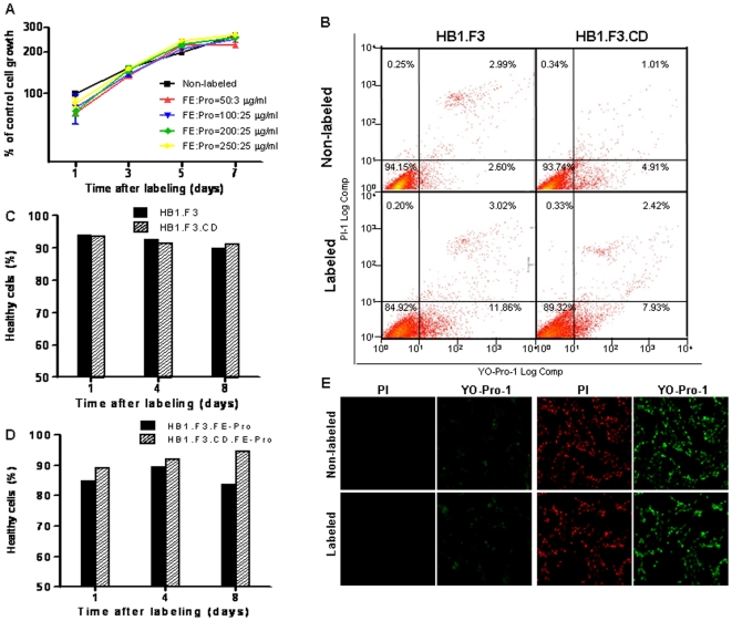
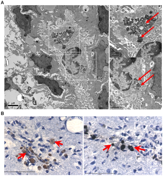
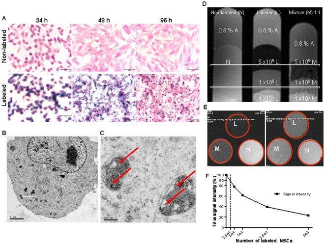
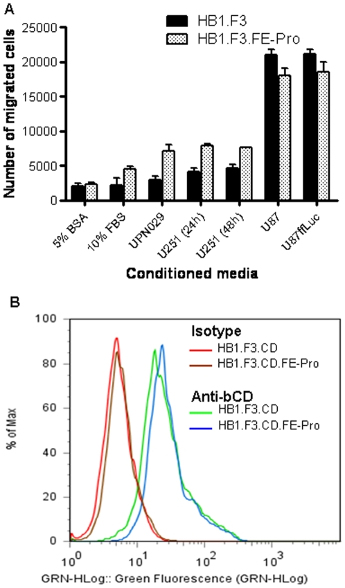
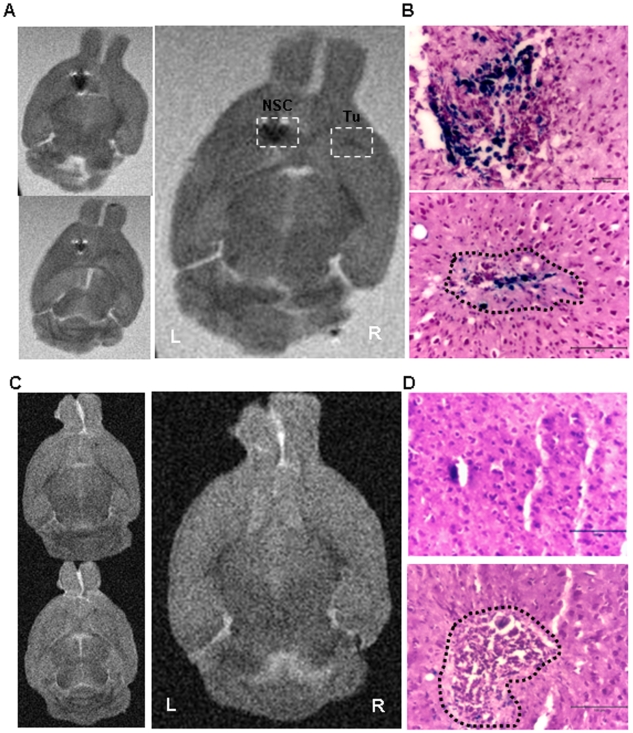
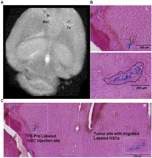
For human use, the characteristics of therapeutic Neural Stem Cells must be clearly defined with any pertubation to the cell including iron labeling requiring reanalysis of cellular physiology. Here, we studied the effect of iron-loading of the therapeutic NSCs, with ferumoxide-protamine sulfate complex (FE-Pro) on viability, proliferation, migratory properties and transgene expression, when compared to non-labeled cells. FE-Pro labeled NSCs were imaged by MRI at tumor sites, after intracranial administration into the hemisphere contralateral to the tumor, in an orthotopic human glioma xenograft mouse model.
FE-Pro labeled NSCs retain their proliferative status, tumor tropism, and maintain stem cell character, while allowing in vivo cellular MRI tracking at 7 Tesla, to monitor their real-time migration and distribution at brain tumor sites. Of significance, this work directly supports the use of FE-Pro-labeled NSCs for real-time tracking in the clinical trial under development: "A Pilot Feasibility Study of Oral 5-Fluorocytosine and Genetically modified Neural Stem Cells Expressing Escherichia coli Cytosine Deaminase for Treatment of Recurrent High-Grade Gliomas".
对于高度侵袭性脑肿瘤胶质母细胞瘤多形性,需要针对已经侵入周围大脑的细胞进行特异性靶向治疗。神经干细胞(NSC)对原发性和侵袭性肿瘤灶的固有肿瘤趋向性,可以被利用来将治疗药物递送到人类侵袭性脑肿瘤细胞中。使用通过永生化治疗性 NSC 系传递的治疗性转基因将前药转化为药物的策略已在动物模型中显示出疗效。因此,治疗性 NSC 被提议用于人类脑肿瘤临床试验。在基于 NSC 的治疗的背景下,MRI 可用于非侵入性地跟踪 NSC 肿瘤靶向的动态时空模式,从而优化治疗策略,并评估治疗的疗效。细胞的铁标记使其能够通过 MRI 可视化和跟踪。因此,我们旨在使用一种可能获得美国食品和药物管理局(FDA)批准的方法,在不影响细胞生理学的情况下对治疗性 NSC 进行铁标记。
对于人类使用,必须清楚地定义治疗性神经干细胞的特性,任何对细胞的干扰,包括铁标记,都需要重新分析细胞生理学。在这里,我们研究了铁负载治疗性 NSCs 的效果,与 ferumoxide-鱼精蛋白硫酸盐复合物(FE-Pro)相比,对非标记细胞的活力、增殖、迁移特性和转基因表达的影响。FE-Pro 标记的 NSCs 在颅内给药到肿瘤对侧半球后,在原位人胶质母细胞瘤异种移植小鼠模型中,通过 MRI 在肿瘤部位成像。
FE-Pro 标记的 NSCs 保留其增殖状态、肿瘤趋向性,并保持干细胞特征,同时允许在体内进行细胞 MRI 跟踪,以监测其在脑肿瘤部位的实时迁移和分布。重要的是,这项工作直接支持了在正在开发的临床试验中使用 FE-Pro 标记的 NSCs 进行实时跟踪:“口服 5-氟胞嘧啶和表达大肠杆菌胞嘧啶脱氨酶的基因修饰神经干细胞治疗复发性高级别神经胶质瘤的初步可行性研究”。