Department of Optometry & Vision Sciences, University of Melbourne, Carlton, Vic. 3053, Australia.
Eur J Neurosci. 2009 Oct;30(8):1517-26. doi: 10.1111/j.1460-9568.2009.06939.x. Epub 2009 Oct 12.
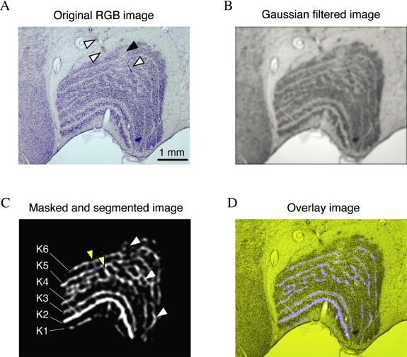
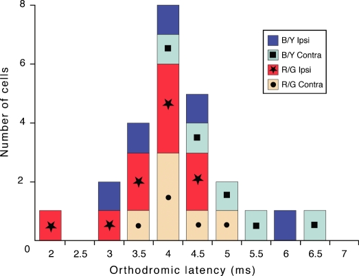
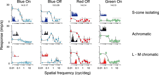
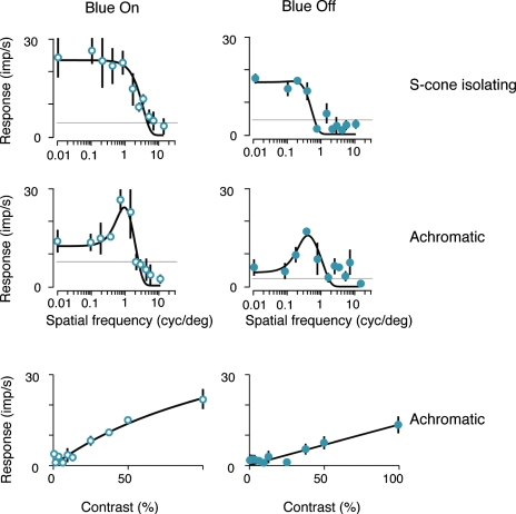
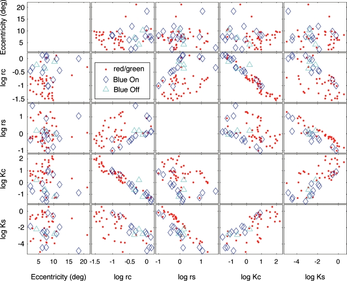
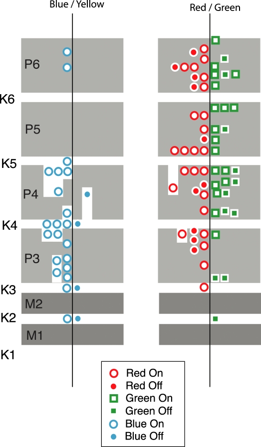
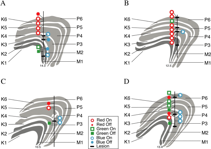
An important problem in the study of the mammalian visual system is whether functionally different retinal ganglion cell types are anatomically segregated further up along the central visual pathway. It was previously demonstrated that, in a New World diurnal monkey (marmoset), the neurones carrying signals from the short-wavelength-sensitive (S) cones [blue-yellow (B/Y)-opponent cells] are predominantly located in the koniocellular layers of the dorsal lateral geniculate nucleus (LGN), whereas the red-green (R/G)-opponent cells carrying signals from the medium- and long-wavelength-sensitive cones are segregated in the parvocellular layers. Here, we used extracellular single-unit recordings followed by histological reconstruction to investigate the distribution of color-selective cells in the LGN of the macaque, an Old World diurnal monkey. Cells were classified using cone-isolating stimuli to identify their cone inputs. Our results indicate that the majority of cells carrying signals from S-cones are located either in the koniocellular layers or in the 'koniocellular bridges' that fully or partially span the parvocellular layers. By contrast, the R/G-opponent cells are located in the parvocellular layers. We conclude that anatomical segregation of B/Y- and R/G-opponent afferent signals for color vision is common to the LGNs of New World and Old World diurnal monkeys.
哺乳动物视觉系统研究中的一个重要问题是,功能不同的视网膜神经节细胞类型在中央视觉通路上是否进一步分离。先前的研究表明,在一种新热带日行动物(狨猴)中,携带来自短波长敏感(S)视锥细胞(蓝-黄(B/Y)-拮抗细胞)信号的神经元主要位于背外侧膝状体核(LGN)的 koniocellular 层中,而携带来自中长波长敏感视锥细胞信号的红-绿(R/G)-拮抗细胞则分离在小细胞层中。在这里,我们使用细胞外单单元记录结合组织学重建,研究了旧世界日行动物猕猴 LGN 中对颜色敏感的细胞的分布。使用分离视锥细胞的刺激来对细胞进行分类,以确定它们的视锥细胞输入。我们的结果表明,携带 S-视锥细胞信号的大多数细胞位于 koniocellular 层或完全或部分跨越小细胞层的“konio 细胞桥”中。相比之下,R/G-拮抗细胞位于小细胞层中。我们得出结论,B/Y-和 R/G-拮抗传入信号的解剖分离对于颜色视觉是新热带和旧世界日行动物 LGN 共有的。