Medical College of Georgia, Department of Physiology, 1120 Fifteenth St, CA-2095, Augusta, GA 30912-3000, USA.
Hypertension. 2010 Jan;55(1):172-9. doi: 10.1161/HYPERTENSIONAHA.109.140459. Epub 2009 Nov 9.
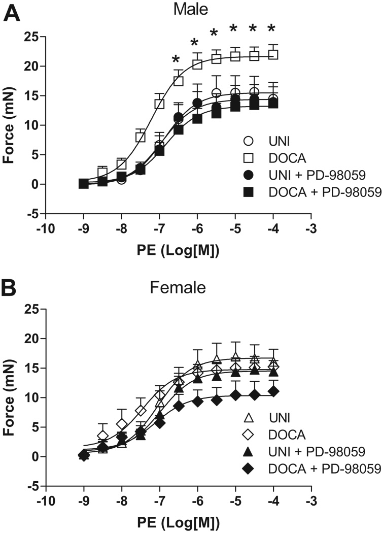
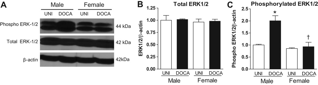
Extracellular signal-regulated kinase (ERK)1/2 has been reported to play a role in vascular dysfunction associated with mineralocorticoid hypertension. We hypothesized that, compared with female rats, an upregulation of ERK1/2 signaling in the vasculature of male rats contributes to augmented contractile responses in mineralocorticoid hypertension. Uninephrectomized male and female Sprague-Dawley rats received desoxycorticosterone acetate (DOCA) pellets (200 mg per animal) and saline to drink for 3 weeks. Control uninephrectomized rats received tap water to drink. Blood pressure, measured by telemetry, was significantly higher in male DOCA rats (191+/-3 mm Hg) compared with female DOCA rats (172+/-7 mm Hg; n=5). DOCA treatment resulted in augmented contractile responses to phenylephrine in aorta (22+/-3 mN; n=6) and small mesenteric arteries (13+/-2 mN; n=6) from male DOCA rats versus uninephrectomized male rats (16+/-3 and 10+/-2 mN, respectively; P<0.05) and female DOCA rats (15+/-1 and 11+/-1 mN, respectively). ERK1/2 inhibition with PD-98059 (10 micromol/L) abrogated increased contraction to phenylephrine in aorta (14+/-2 mN) and small mesenteric arteries (10+/-2 mN) from male DOCA rats, without any effects in arteries from male uninephrectomized or female animals. Compared with the other groups, phosphorylated ERK1/2 levels were increased in the aorta from male DOCA rats, whereas mitogen-activated protein kinase phosphatase 1 expression was decreased. Interleukin-10 plasma levels, which positively regulate mitogen-activated protein kinase phosphatase 1 activity, were reduced in male DOCA-salt rats. We speculate that augmented vascular reactivity in male hypertensive rats is mediated via activation of the ERK1/2 pathway. In addition, mitogen-activated protein kinase phosphatase 1 and interleukin 10 play regulatory roles in this process.
细胞外信号调节激酶(ERK)1/2 已被报道在与醛固酮高血压相关的血管功能障碍中发挥作用。我们假设,与雌性大鼠相比,雄性大鼠血管中 ERK1/2 信号的上调导致醛固酮高血压中收缩反应增强。单侧肾切除的雄性和雌性 Sprague-Dawley 大鼠接受去氧皮质酮乙酸酯(DOCA)丸(每只动物 200mg)和盐水饮用 3 周。对照单侧肾切除大鼠饮用自来水。通过遥测测量的血压在雄性 DOCA 大鼠(191+/-3mmHg)中显著高于雌性 DOCA 大鼠(172+/-7mmHg;n=5)。DOCA 处理导致来自雄性 DOCA 大鼠的主动脉(22+/-3mN;n=6)和小肠系膜动脉(13+/-2mN;n=6)对苯肾上腺素的收缩反应增强,而与单侧肾切除的雄性大鼠相比(分别为 16+/-3 和 10+/-2mN;P<0.05)和雌性 DOCA 大鼠(15+/-1 和 11+/-1mN,分别)。PD-98059(10µmol/L)抑制 ERK1/2 可消除来自雄性 DOCA 大鼠的主动脉(14+/-2mN)和小肠系膜动脉(10+/-2mN)对苯肾上腺素的收缩增加,而对来自雄性单侧肾切除或雌性动物的动脉没有任何作用。与其他组相比,雄性 DOCA 大鼠主动脉中的磷酸化 ERK1/2 水平增加,而丝裂原活化蛋白激酶磷酸酶 1 的表达减少。白细胞介素-10 血浆水平降低,白细胞介素-10 正向调节丝裂原活化蛋白激酶磷酸酶 1 的活性,在雄性 DOCA-盐大鼠中降低。我们推测,雄性高血压大鼠血管反应性增强是通过 ERK1/2 途径的激活介导的。此外,丝裂原活化蛋白激酶磷酸酶 1 和白细胞介素 10 在这个过程中起调节作用。