Wagner Claudia C, Bauer Martin, Karch Rudolf, Feurstein Thomas, Kopp Stephan, Chiba Peter, Kletter Kurt, Löscher Wolfgang, Müller Markus, Zeitlinger Markus, Langer Oliver
Department of Clinical Pharmacology, Medical University of Vienna, Vienna, Austria.
J Nucl Med. 2009 Dec;50(12):1954-61. doi: 10.2967/jnumed.109.063289. Epub 2009 Nov 12.
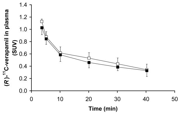
Tariquidar, a potent, nontoxic, third-generation P-glycoprotein (P-gp) inhibitor, is a possible reversal agent for central nervous system drug resistance. In animal studies, tariquidar has been shown to increase the delivery of P-gp substrates into the brain by severalfold. The aim of this study was to measure P-gp function at the human blood-brain barrier (BBB) after tariquidar administration using PET and the model P-gp substrate (R)-(11)C-verapamil.
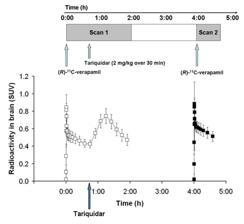
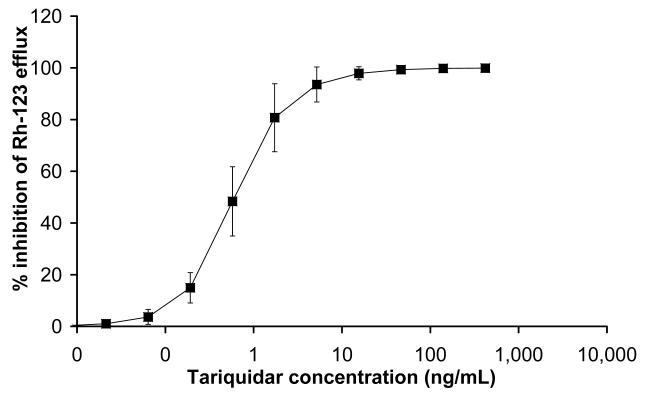
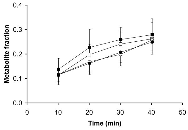
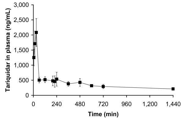
Five healthy volunteers underwent paired (R)-(11)C-verapamil PET scans and arterial blood sampling before and at 2 h 50 min after intravenous administration of tariquidar (2 mg/kg of body weight). The inhibition of P-gp on CD56-positive peripheral lymphocytes of each volunteer was determined by means of the (123)Rh efflux assay. Tariquidar concentrations in venous plasma were quantified using liquid chromatography/mass spectrometry.
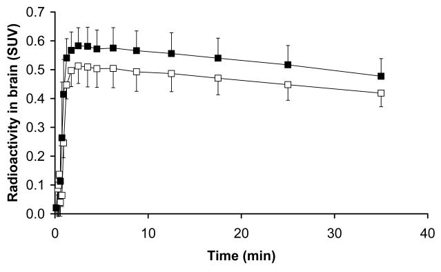
Tariquidar administration resulted in significant increases (Wilcoxon test for paired samples) in the distribution volume (DV, +24% +/- 15%) and influx rate constant (K(1), +49% +/- 36%) of (R)-(11)C-verapamil across the BBB (DV, 0.65 +/- 0.13 and 0.80 +/- 0.07, P = 0.043; K(1), 0.034 +/- 0.009 and 0.049 +/- 0.009, P = 0.043, before and after tariquidar administration, respectively). A strong correlation was observed between the change in brain DV after administration of tariquidar and tariquidar exposure in plasma (r = 0.90, P = 0.037). The mean plasma concentration of tariquidar achieved during the second PET scan (490 +/- 166 ng/mL) corresponded to 100% inhibition of P-gp function in peripheral lymphocytes.
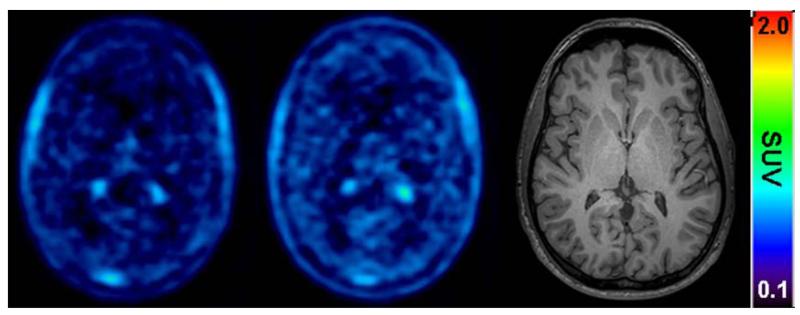
Tariquidar significantly increased brain penetration of (R)-(11)C-verapamil-derived activity due to increased influx. As opposed to peripheral P-gp function, central P-gp inhibition appeared to be far from complete after the administered tariquidar dose.
塔里喹达是一种强效、无毒的第三代P-糖蛋白(P-gp)抑制剂,可能是中枢神经系统耐药的逆转剂。在动物研究中,塔里喹达已被证明可使P-gp底物进入大脑的量增加数倍。本研究的目的是使用正电子发射断层扫描(PET)和模型P-gp底物(R)-(11)C-维拉帕米,测量塔里喹达给药后人体血脑屏障(BBB)处的P-gp功能。
5名健康志愿者在静脉注射塔里喹达(2mg/kg体重)前及给药后2小时50分钟接受配对的(R)-(11)C-维拉帕米PET扫描和动脉血采样。通过(123)铑流出试验测定每名志愿者CD56阳性外周淋巴细胞上P-gp的抑制情况。使用液相色谱/质谱法定量静脉血浆中塔里喹达的浓度。
给予塔里喹达后,(R)-(11)C-维拉帕米穿过BBB的分布容积(DV,+24%±15%)和流入速率常数(K1,+49%±36%)显著增加(配对样本的Wilcoxon检验)(给药前和给药后DV分别为0.65±0.13和0.80±0.07,P = 0.043;K1分别为0.034±0.009和0.049±0.009,P = 0.043)。观察到给予塔里喹达后大脑DV的变化与血浆中塔里喹达的暴露量之间存在强相关性(r = 0.90,P = 0.037)。第二次PET扫描期间达到的塔里喹达平均血浆浓度(490±166ng/mL)相当于外周淋巴细胞中P-gp功能的100%抑制。
由于流入增加,塔里喹达显著增加了(R)-(11)C-维拉帕米衍生活性的脑渗透。与外周P-gp功能相反,给予塔里喹达剂量后,中枢P-gp抑制似乎远未完全。