Halle Stephan, Dujardin Hélène C, Bakocevic Nadja, Fleige Henrike, Danzer Heike, Willenzon Stefanie, Suezer Yasemin, Hämmerling Günter, Garbi Natalio, Sutter Gerd, Worbs Tim, Förster Reinhold
Institute of Immunology, Hannover Medical School, 30625 Hannover, Germany.
J Exp Med. 2009 Nov 23;206(12):2593-601. doi: 10.1084/jem.20091472. Epub 2009 Nov 16.
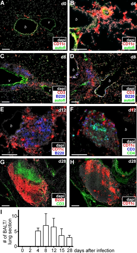
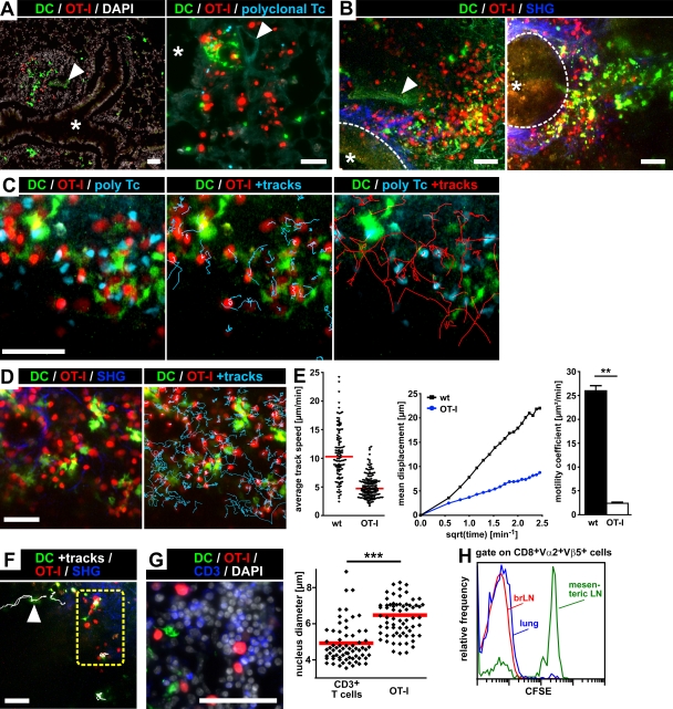
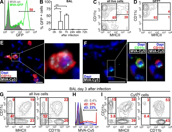
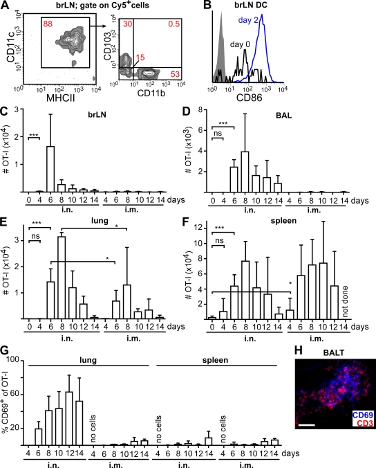
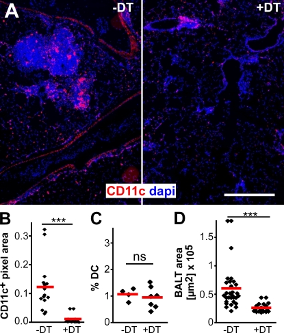
Mucosal vaccination via the respiratory tract can elicit protective immunity in animal infection models, but the underlying mechanisms are still poorly understood. We show that a single intranasal application of the replication-deficient modified vaccinia virus Ankara, which is widely used as a recombinant vaccination vector, results in prominent induction of bronchus-associated lymphoid tissue (BALT). Although initial peribronchiolar infiltrations, characterized by the presence of dendritic cells (DCs) and few lymphocytes, can be found 4 d after virus application, organized lymphoid structures with segregated B and T cell zones are first observed at day 8. After intratracheal application, in vitro-differentiated, antigen-loaded DCs rapidly migrate into preformed BALT and efficiently activate antigen-specific T cells, as revealed by two-photon microscopy. Furthermore, the lung-specific depletion of DCs in mice that express the diphtheria toxin receptor under the control of the CD11c promoter interferes with BALT maintenance. Collectively, these data identify BALT as tertiary lymphoid structures supporting the efficient priming of T cell responses directed against unrelated airborne antigens while crucially requiring DCs for its sustained presence.
通过呼吸道进行黏膜疫苗接种可在动物感染模型中引发保护性免疫,但其潜在机制仍知之甚少。我们发现,单次鼻内接种广泛用作重组疫苗载体的复制缺陷型安卡拉痘苗病毒,会显著诱导支气管相关淋巴组织(BALT)的形成。尽管在接种病毒4天后可发现以树突状细胞(DCs)和少量淋巴细胞为特征的初始支气管周围浸润,但在第8天首次观察到具有分离的B细胞和T细胞区的有组织的淋巴结构。气管内接种后,如双光子显微镜所示,体外分化的、负载抗原的DCs迅速迁移到预先形成的BALT中,并有效激活抗原特异性T细胞。此外,在CD11c启动子控制下表达白喉毒素受体的小鼠中,DCs的肺特异性耗竭会干扰BALT的维持。总体而言,这些数据表明BALT是三级淋巴结构,支持针对无关空气传播抗原的T细胞反应的有效启动,同时其持续存在至关重要地需要DCs。