Department of Anatomy and Neurobiology, School of Medicine, University of California at Irvine, Irvine, CA 92697-1275, USA.
Brain Struct Funct. 2009 Dec;214(1):25-35. doi: 10.1007/s00429-009-0231-7. Epub 2009 Nov 21.
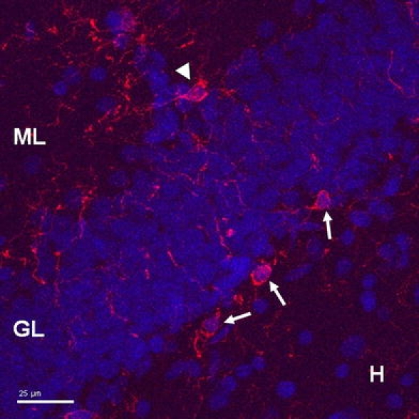
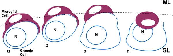
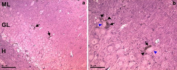
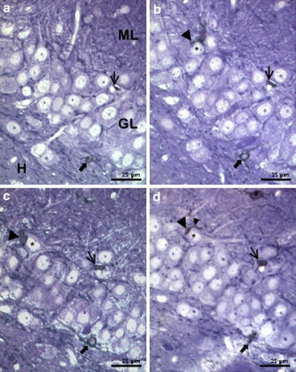
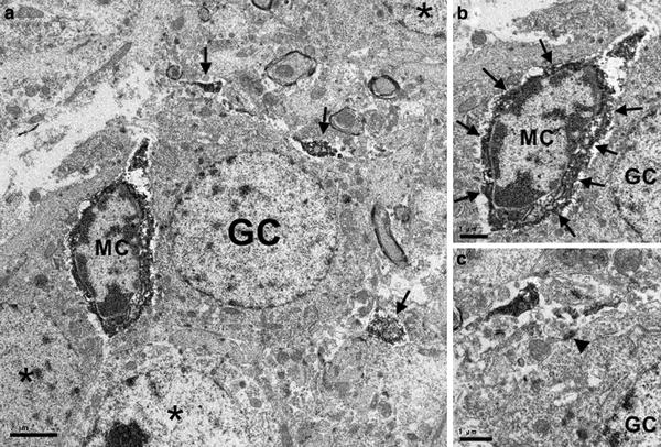
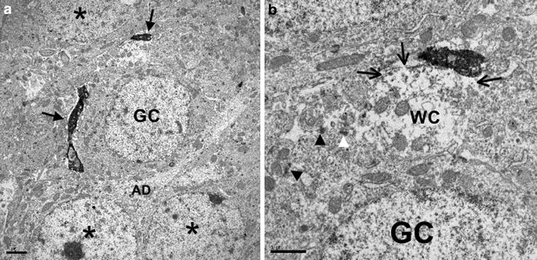
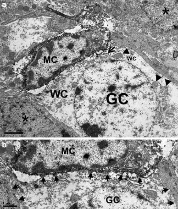
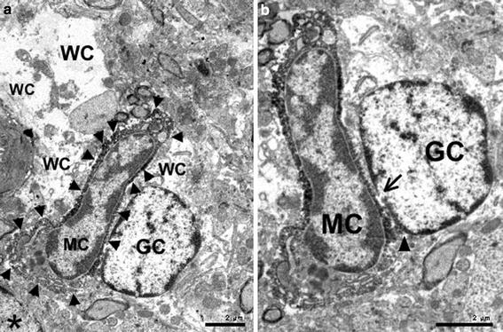
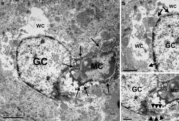
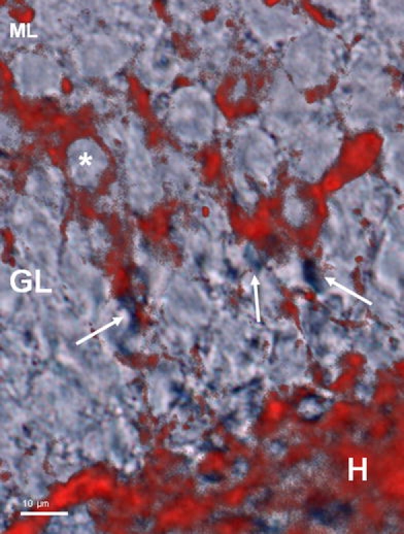
Microglial cells are constantly monitoring the central nervous system for sick or dying cells and pathogens. Previous studies showed that the microglial cells in the dentate gyrus have a heterogeneous morphology with multipolar cells in the hilus and fusiform cells apposed to the granule cell layer both at the hilar and at the molecular layer borders. Although previous studies showed that the microglia in the dentate gyrus were not activated, the data in the present study show dying granule cells apposed by Iba1-immunolabeled microglial cell bodies and their processes both at hilar and at molecular layer borders of the granule cell layer. Initially, these Iba1-labeled microglial cells surround individual, intact granule cell bodies. When small openings in the plasma membrane of granule cells are observed, microglial cells are apposed to these openings. When larger openings in the plasma membrane occur at this site of apposition, the granule cells display watery perikaryal cytoplasm, watery nucleoplasm and damaged organelles. Such morphological features are characteristic of neuronal edema. The data also show that following this localized disintegration of the granule cell's plasma membrane, the Iba1-labeled microglial cell body is found within the electron-lucent perikaryal cytoplasm of the granule cell, where it is adjacent to the granule cell's nucleus which is deformed. We propose that granule cells are dying by a novel microglia-associated mechanism that involves lysis of their plasma membranes followed by neuronal edema and nuclear phagocytosis. Based on the morphological evidence, this type of cell death differs from either apoptosis or necrosis.
小胶质细胞不断监测中枢神经系统中病变或死亡的细胞和病原体。先前的研究表明,齿状回中的小胶质细胞具有异质形态,其在门区有多极细胞,在与颗粒细胞层相邻的门区和分子层边界处有梭形细胞。尽管先前的研究表明齿状回中的小胶质细胞没有被激活,但本研究的数据显示,与 Iba1 免疫标记的小胶质细胞体及其突起相邻的死亡颗粒细胞位于颗粒细胞层的门区和分子层边界。最初,这些 Iba1 标记的小胶质细胞围绕单个完整的颗粒细胞体。当观察到颗粒细胞膜上出现小的开口时,小胶质细胞就会附着在这些开口上。当在这个附着部位出现更大的细胞膜开口时,颗粒细胞显示出水样胞质、水样核质和受损的细胞器。这种形态特征是神经元水肿的特征。这些数据还表明,在颗粒细胞的质膜局部崩解后,Iba1 标记的小胶质细胞体被发现位于颗粒细胞的电子透明胞质内,与颗粒细胞的变形核相邻。我们提出,颗粒细胞通过一种新的与小胶质细胞相关的机制死亡,该机制涉及质膜的裂解,随后是神经元水肿和核吞噬。基于形态学证据,这种类型的细胞死亡不同于凋亡或坏死。