Department of Biochemistry, University of Delhi South Campus, New Delhi, India.
PLoS One. 2009 Nov 25;4(11):e8028. doi: 10.1371/journal.pone.0008028.
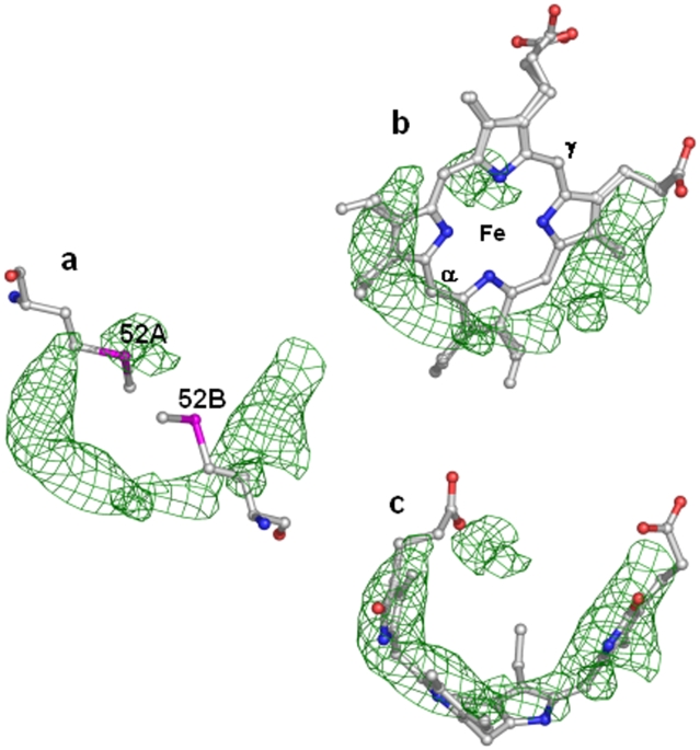
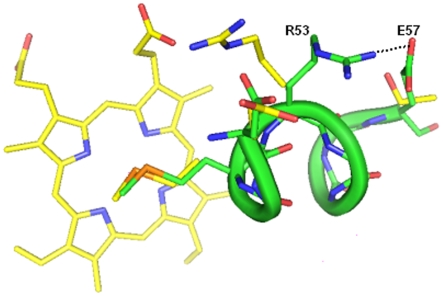
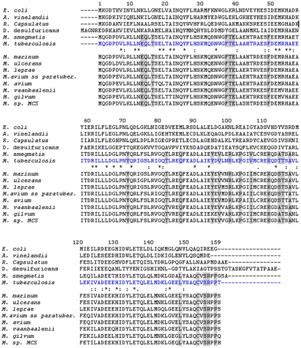
Emergence of tuberculosis as a global health threat has necessitated an urgent search for new antitubercular drugs entailing determination of 3-dimensional structures of a large number of mycobacterial proteins for structure-based drug design. The essential requirement of ferritins/bacterioferritins (proteins involved in iron storage and homeostasis) for the survival of several prokaryotic pathogens makes these proteins very attractive targets for structure determination and inhibitor design. Bacterioferritins (Bfrs) differ from ferritins in that they have additional noncovalently bound haem groups. The physiological role of haem in Bfrs is not very clear but studies indicate that the haem group is involved in mediating release of iron from Bfr by facilitating reduction of the iron core. To further enhance our understanding, we have determined the crystal structure of the selenomethionyl analog of bacterioferritin A (SeMet-BfrA) from Mycobacterium tuberculosis (Mtb). Unexpectedly, electron density observed in the crystals of SeMet-BfrA analogous to haem location in bacterioferritins, shows a demetallated and degraded product of haem. This unanticipated observation is a consequence of the altered spatial electronic environment around the axial ligands of haem (in lieu of Met52 modification to SeMet52). Furthermore, the structure of Mtb SeMet-BfrA displays a possible lost protein interaction with haem propionates due to formation of a salt bridge between Arg53-Glu57, which appears to be unique to Mtb BfrA, resulting in slight modulation of haem binding pocket in this organism. The crystal structure of Mtb SeMet-BfrA provides novel leads to physiological function of haem in Bfrs. If validated as a drug target, it may also serve as a scaffold for designing specific inhibitors. In addition, this study provides evidence against the general belief that a selenium derivative of a protein represents its true physiological native structure.
结核分枝杆菌作为一种全球健康威胁的出现,促使人们迫切需要寻找新的抗结核药物,这需要确定大量分枝杆菌蛋白的三维结构,以便进行基于结构的药物设计。铁蛋白/菌铁蛋白(参与铁储存和体内平衡的蛋白质)对于许多原核病原体的生存至关重要,这使得这些蛋白质成为确定结构和抑制剂设计的极具吸引力的靶标。菌铁蛋白(Bfrs)与铁蛋白的不同之处在于它们具有额外的非共价结合的血红素基团。血红素在 Bfrs 中的生理作用尚不清楚,但研究表明,血红素基团参与通过促进铁核的还原来介导从 Bfr 中释放铁。为了进一步增强我们的理解,我们已经确定了结核分枝杆菌(Mtb)菌铁蛋白 A(SeMet-BfrA)的硒代甲硫氨酸类似物的晶体结构。出乎意料的是,在 SeMet-BfrA 的晶体中观察到的电子密度类似于菌铁蛋白中血红素的位置,显示出血红素的脱金属和降解产物。这种意外的观察结果是由于血红素轴向配体周围的空间电子环境发生改变(代替 Met52 修饰为 SeMet52)的结果。此外,Mtb SeMet-BfrA 的结构显示出与血红素丙酸盐可能失去的蛋白质相互作用,由于 Arg53-Glu57 之间形成盐桥,这似乎是 Mtb BfrA 所特有的,导致在该生物体中血红素结合口袋略有调制。Mtb SeMet-BfrA 的晶体结构为血红素在 Bfrs 中的生理功能提供了新的线索。如果被验证为药物靶标,它也可以作为设计特定抑制剂的支架。此外,这项研究提供了证据,证明了一般的信念,即蛋白质的硒衍生物代表其真实的生理天然结构是错误的。