Department of Biosciences, New England College of Optometry, Boston, MA 02115, USA.
Prog Retin Eye Res. 2010 Mar;29(2):144-68. doi: 10.1016/j.preteyeres.2009.12.002. Epub 2009 Dec 29.
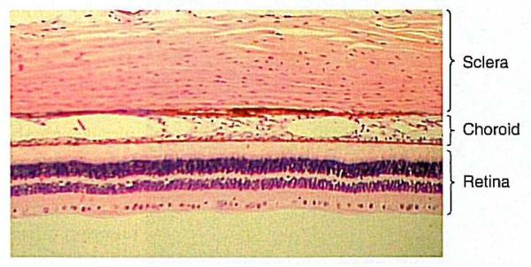
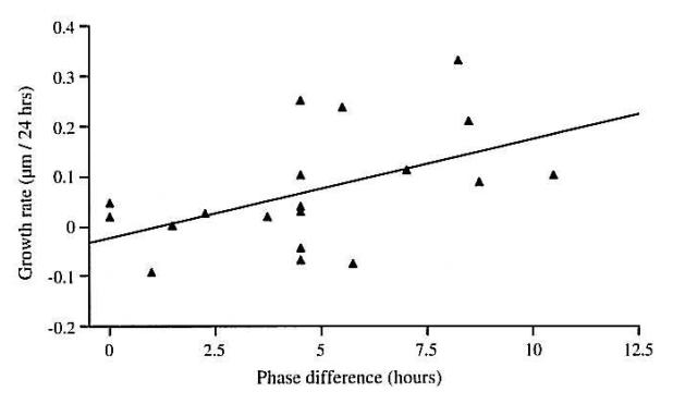
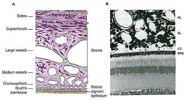
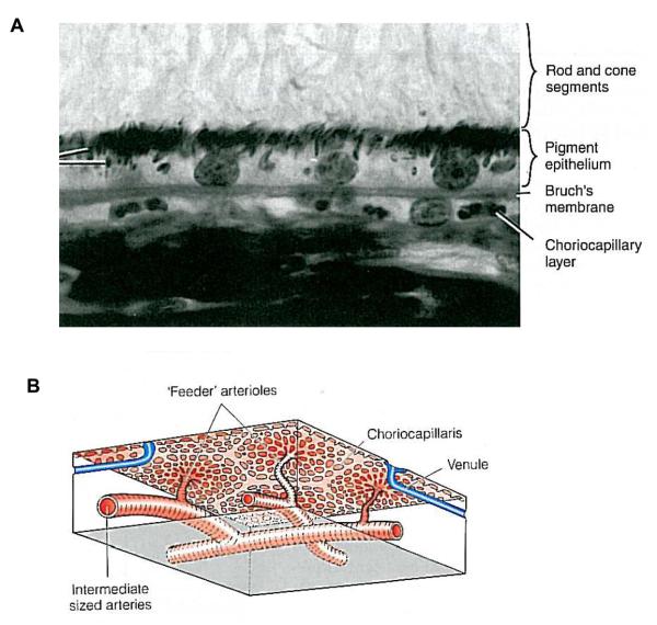
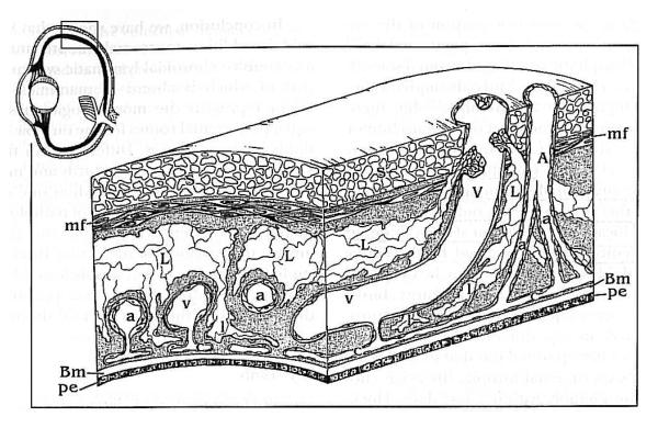
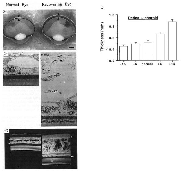
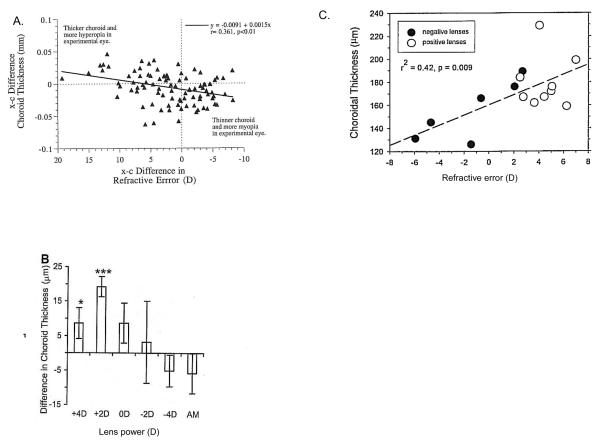
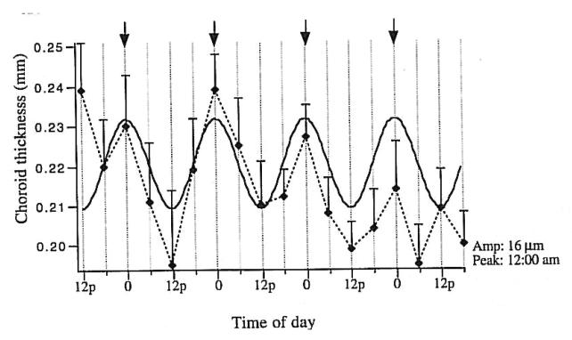
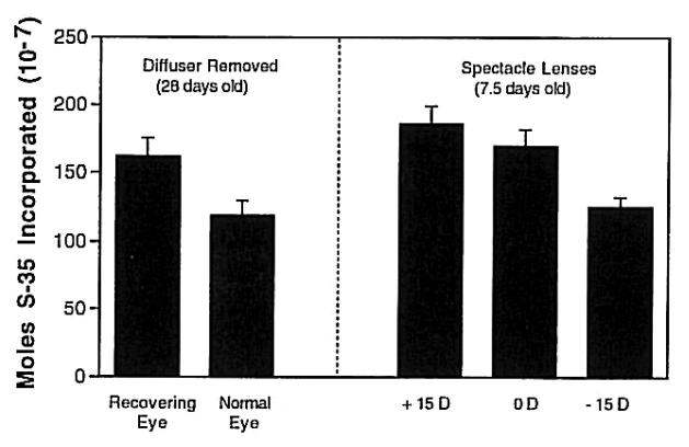
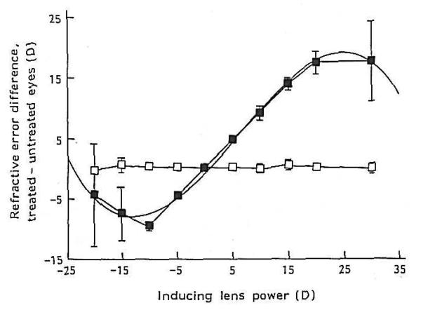
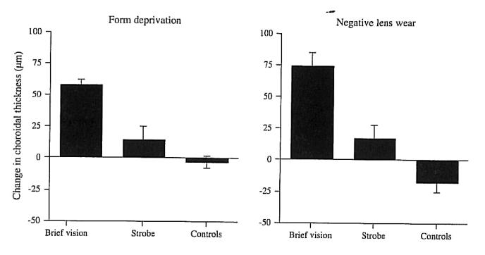
The choroid of the eye is primarily a vascular structure supplying the outer retina. It has several unusual features: It contains large membrane-lined lacunae, which, at least in birds, function as part of the lymphatic drainage of the eye and which can change their volume dramatically, thereby changing the thickness of the choroid as much as four-fold over a few days (much less in primates). It contains non-vascular smooth muscle cells, especially behind the fovea, the contraction of which may thin the choroid, thereby opposing the thickening caused by expansion of the lacunae. It has intrinsic choroidal neurons, also mostly behind the central retina, which may control these muscles and may modulate choroidal blood flow as well. These neurons receive sympathetic, parasympathetic and nitrergic innervation. The choroid has several functions: Its vasculature is the major supply for the outer retina; impairment of the flow of oxygen from choroid to retina may cause Age-Related Macular Degeneration. The choroidal blood flow, which is as great as in any other organ, may also cool and warm the retina. In addition to its vascular functions, the choroid contains secretory cells, probably involved in modulation of vascularization and in growth of the sclera. Finally, the dramatic changes in choroidal thickness move the retina forward and back, bringing the photoreceptors into the plane of focus, a function demonstrated by the thinning of the choroid that occurs when the focal plane is moved back by the wearing of negative lenses, and, conversely, by the thickening that occurs when positive lenses are worn. In addition to focusing the eye, more slowly than accommodation and more quickly than emmetropization, we argue that the choroidal thickness changes also are correlated with changes in the growth of the sclera, and hence of the eye. Because transient increases in choroidal thickness are followed by a prolonged decrease in synthesis of extracellular matrix molecules and a slowing of ocular elongation, and attempts to decouple the choroidal and scleral changes have largely failed, it seems that the thickening of the choroid may be mechanistically linked to the scleral synthesis of macromolecules, and thus may play an important role in the homeostatic control of eye growth, and, consequently, in the etiology of myopia and hyperopia.
眼睛的脉络膜主要是为外视网膜提供营养的血管结构。它具有几个不寻常的特征:它包含大的膜衬腔隙,至少在鸟类中,这些腔隙作为眼睛淋巴引流的一部分起作用,可以显著改变其体积,从而使脉络膜的厚度在几天内变化高达四倍(在灵长类动物中则小得多)。它含有非血管平滑肌细胞,特别是在黄斑后面,其收缩可能使脉络膜变薄,从而抵消由腔隙扩张引起的增厚。它具有内在的脉络膜神经元,也主要位于中央视网膜后面,这些神经元可能控制这些肌肉,并调节脉络膜的血流。这些神经元接收交感神经、副交感神经和硝基能神经支配。脉络膜具有多种功能:其血管系统是外视网膜的主要供应者;脉络膜向视网膜供氧的流动受损可能导致年龄相关性黄斑变性。脉络膜血流与任何其他器官一样大,也可以冷却和温暖视网膜。除了其血管功能外,脉络膜还含有分泌细胞,可能参与血管生成和巩膜的生长调节。最后,脉络膜厚度的显著变化使视网膜前后移动,使光感受器进入焦点平面,这一功能通过佩戴负透镜使焦点平面向后移动时脉络膜变薄来证明,反之,通过佩戴正透镜使脉络膜变厚来证明。除了聚焦眼睛外,比调节更慢,比正视化更快,我们认为脉络膜厚度的变化也与巩膜的生长变化有关,因此也与眼睛有关。由于脉络膜厚度的短暂增加后伴随着细胞外基质分子合成的延长减少和眼球伸长的减缓,并且试图使脉络膜和巩膜的变化解耦的尝试基本上都失败了,因此脉络膜的增厚似乎在机械上与巩膜的大分子合成有关,因此可能在眼球生长的稳态控制中发挥重要作用,从而在近视和远视的病因中发挥重要作用。