Institute of Neuroscience, Newcastle University, Newcastle upon Tyne, UK.
Neuroimage. 2010 Apr 15;50(3):1099-108. doi: 10.1016/j.neuroimage.2009.12.103. Epub 2010 Jan 4.
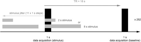
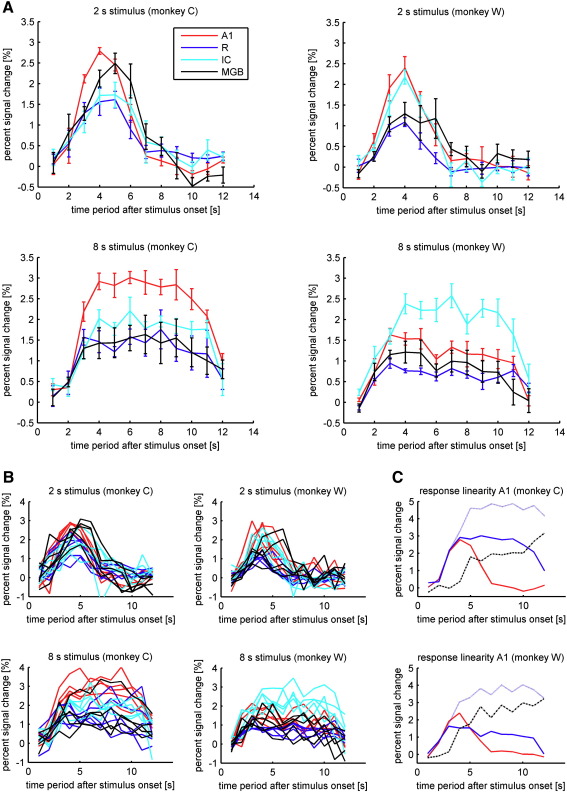
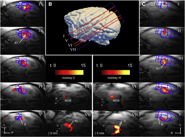
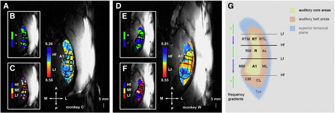
Non-human-primate fMRI is becoming increasingly recognised as the missing link between the widely applied methods of human imaging and intracortical animal electrophysiology. A crucial requirement for the optimal application of this method is the precise knowledge of the time course of the Blood Oxygenation Level Dependent (BOLD) signal. We mapped the BOLD signal time course in the inferior colliculus (IC), medial geniculate body (MGB) and in tonotopically defined fields in the auditory cortex of two macaques. The results show little differences in the BOLD-signal time courses within the auditory pathway. However, we observed systematic differences in the magnitude of the change in the BOLD signal with significantly stronger signal changes in field A1 of the auditory cortex compared to field R. The measured time course of the signal was in good agreement with similar studies in human auditory cortex but showed considerable differences to data reported from macaque visual cortex. Consistent with the studies in humans we measured a peak in the BOLD response around 4 s after the onset of 2-s broadband noise stimuli while previous studies recorded from the primary visual cortex of the same species reported the earliest peaks to short visual stimuli several seconds later. The comparison of our results with previous studies does not support differences in haemodynamic responses within the auditory system between human and non-human primates. Furthermore, the data will aid optimal design of future auditory fMRI studies in non-human primates.
非灵长类动物 fMRI 正逐渐被认为是广泛应用的人类成像方法和皮质内动物电生理学之间缺失的一环。这种方法的最佳应用的一个关键要求是精确了解血氧水平依赖 (BOLD) 信号的时间过程。我们在两只猕猴的下丘 (IC)、内侧膝状体 (MGB) 和听觉皮层的音调定义区域中绘制了 BOLD 信号的时间过程。结果表明,听觉通路上的 BOLD 信号时间过程差异很小。然而,我们观察到 BOLD 信号变化幅度的系统差异,与听觉皮层的 A1 区相比,R 区的信号变化明显更强。测量的信号时间过程与人听觉皮层的类似研究非常吻合,但与来自猕猴视觉皮层的数据报告有很大差异。与人类的研究一致,我们在 2 秒宽带噪声刺激开始后约 4 秒测量到 BOLD 反应的峰值,而之前同一物种的初级视觉皮层的研究记录到的最早峰值是几秒钟后短视觉刺激。我们的结果与之前的研究比较不支持人类和非人类灵长类动物听觉系统内血液动力学反应的差异。此外,这些数据将有助于未来非人类灵长类动物听觉 fMRI 研究的最佳设计。