Department of Radiology, University of Pennsylvania, Philadelphia, PA 19104-6389, USA.
Neuroimage. 2010 Apr 15;50(3):1004-16. doi: 10.1016/j.neuroimage.2010.01.041. Epub 2010 Jan 18.
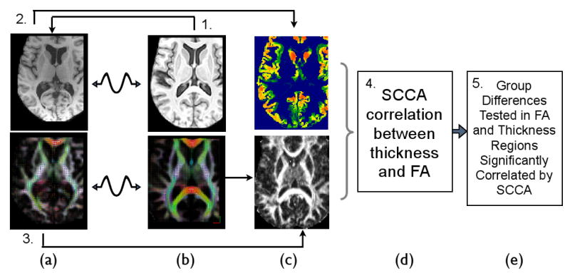
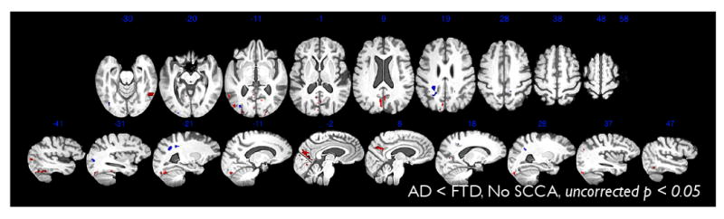
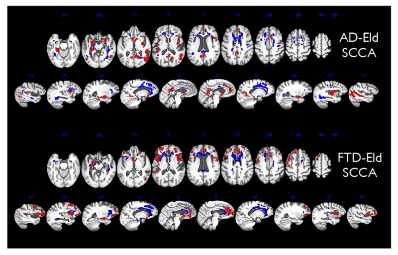
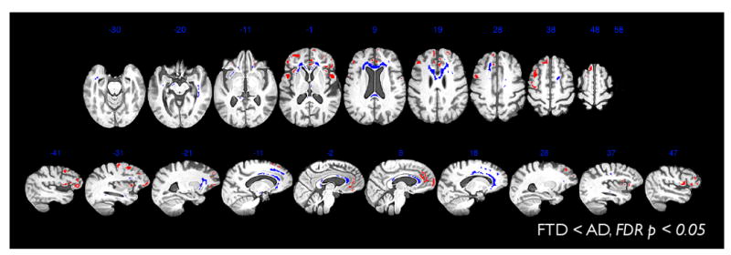
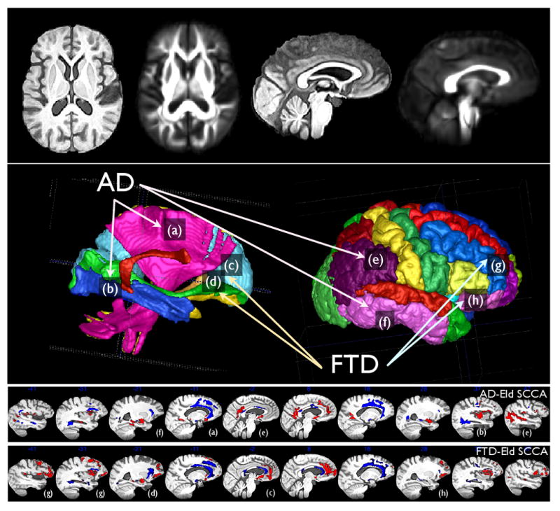
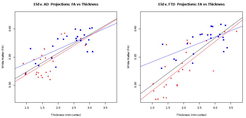
We use a new, unsupervised multivariate imaging and analysis strategy to identify related patterns of reduced white matter integrity, measured with the fractional anisotropy (FA) derived from diffusion tensor imaging (DTI), and decreases in cortical thickness, measured by high resolution T1-weighted imaging, in Alzheimer's disease (AD) and frontotemporal dementia (FTD). This process is based on a novel computational model derived from sparse canonical correlation analysis (SCCA) that allows us to automatically identify mutually predictive, distributed neuroanatomical regions from different imaging modalities. We apply the SCCA model to a dataset that includes 23 control subjects that are demographically matched to 49 subjects with autopsy or CSF-biomarker-diagnosed AD (n=24) and FTD (n=25) with both DTI and T1-weighted structural imaging. SCCA shows that the FTD-related frontal and temporal degeneration pattern is correlated across modalities with permutation corrected p<0.0005. In AD, we find significant association between cortical thinning and reduction in white matter integrity within a distributed parietal and temporal network (p<0.0005). Furthermore, we show that-within SCCA identified regions-significant differences exist between FTD and AD cortical-connective degeneration patterns. We validate these distinct, multimodal imaging patterns by showing unique relationships with cognitive measures in AD and FTD. We conclude that SCCA is a potentially valuable approach in image analysis that can be applied productively to distinguishing between neurodegenerative conditions.
我们使用一种新的、无监督的多变量成像和分析策略,来识别阿尔茨海默病(AD)和额颞叶痴呆(FTD)中,与白质完整性降低相关的模式,这种降低是通过弥散张量成像(DTI)得出的分数各向异性(FA)来测量的,以及皮质厚度的降低,通过高分辨率 T1 加权成像来测量。这个过程是基于一个新的从稀疏典型相关分析(SCCA)得出的计算模型,它使我们能够自动从不同的成像模式中识别出相互预测的、分布的神经解剖区域。我们将 SCCA 模型应用于一个数据集,其中包括 23 名在人口统计学上与 49 名尸检或 CSF 生物标志物诊断为 AD(n=24)和 FTD(n=25)的受试者匹配的对照受试者,这些受试者均具有 DTI 和 T1 加权结构成像。SCCA 显示,FTD 相关的额颞叶变性模式在模态之间具有相关性,经过置换校正后 p<0.0005。在 AD 中,我们发现皮质变薄与皮质下和颞叶网络内的白质完整性降低之间存在显著的关联(p<0.0005)。此外,我们还表明,在 SCCA 确定的区域内,FTD 和 AD 的皮质连接退化模式之间存在显著差异。我们通过在 AD 和 FTD 中显示与认知测量的独特关系,验证了这些独特的、多模态的成像模式。我们的结论是,SCCA 是一种有潜力的图像分析方法,可用于有效地区分神经退行性疾病。