Cellular and Molecular Imaging Laboratory, Department of Radiology, Henry Ford Hospital, Detroit, Michigan, United States of America.
PLoS One. 2010 Jan 15;5(1):e8727. doi: 10.1371/journal.pone.0008727.
Anti-angiogenic treatments of malignant tumors targeting vascular endothelial growth factor receptors (VEGFR) tyrosine kinase are being used in different early stages of clinical trials. Very recently, VEGFR tyrosine kinase inhibitor (Vetanalib, PTK787) was used in glioma patient in conjunction with chemotherapy and radiotherapy. However, changes in the tumor size, tumor vascular permeability, vascular density, expression of VEGFR2 and other angiogenic factors in response to PTK787 are not well documented. This study was to determine the changes in tumor size, vascular permeability, fractional plasma volume and expression of VEGFR2 in PTK787 treated U-251 glioma rat model by in vivo magnetic resonance imaging (MRI) and single photon emission computed tomography (SPECT). The findings were validated with histochemical and western blot studies.
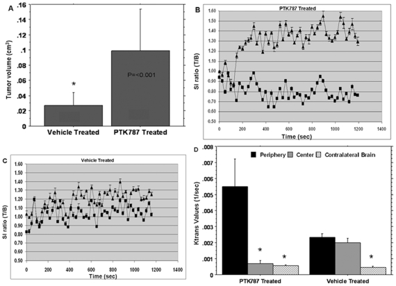
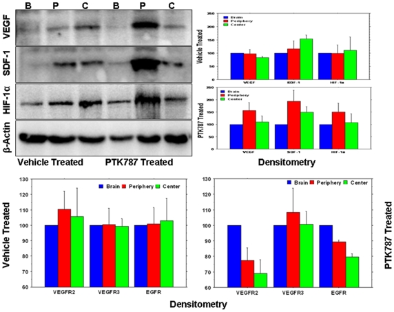
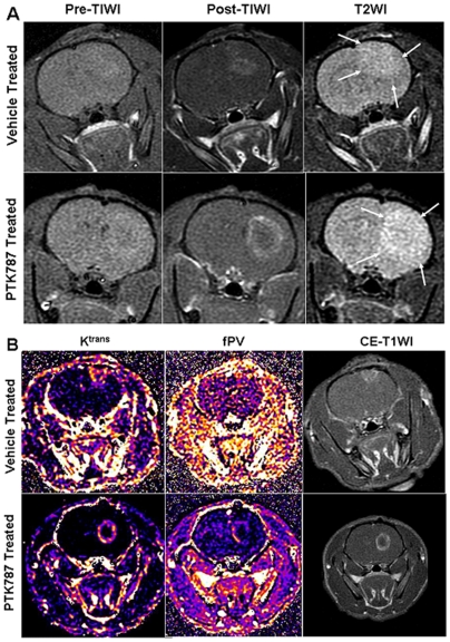
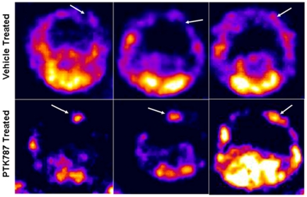
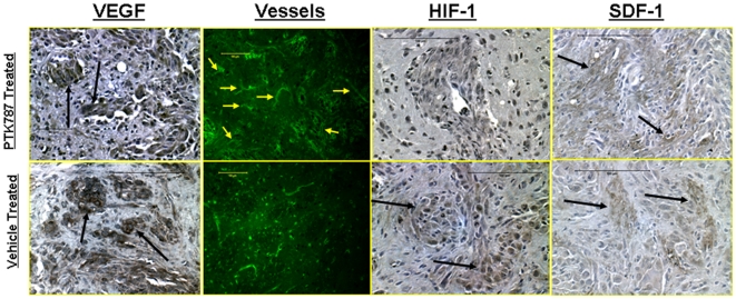
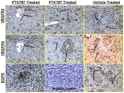
Seven days after implantation of U251 glioma cells, animals were treated with either PTK787 or vehicle-only for two weeks, and then tumor size, tumor vascular permeability transfer constant (K(trans)), fractional plasma volume (fPV) and expression of VEGFR2 and other relevant angiogenic factors were assessed by in vivo MRI and SPECT (Tc-99-HYNIC-VEGF), and by immunohistochemistry and western blot analysis. Dynamic contrast-enhanced MRI (DCE-MRI) using a high molecular weight contrast agent albumin-(GdDTPA) showed significantly increased K(trans) at the rim of the treated tumors compared to that of the central part of the treated as well as the untreated (vehicle treated) tumors. Size of the tumors was also increased in the treated group. Expression of VEGFR2 detected by Tc-99m-HYNIC-VEGF SPECT also showed significantly increased activity in the treated tumors. In PTK787-treated tumors, histological staining revealed increase in microvessel density in the close proximity to the tumor border. Western blot analysis indicated increased expression of VEGF, SDF-1, HIF-1alpha, VEGFR2, VEGFR3 and EGFR at the peripheral part of the treated tumors compared to that of central part of the treated tumors. Similar expression patters were not observed in vehicle treated tumors.
These findings indicate that PTK787 treatment induced over expression of VEGF as well as the Flk-1/VEGFR2 receptor tyrosine kinase, especially at the rim of the tumor, as proven by DCE-MRI, SPECT imaging, immunohistochemistry and western blot.
针对血管内皮生长因子受体(VEGFR)酪氨酸激酶的抗血管生成治疗药物已被用于恶性肿瘤的不同临床试验早期阶段。最近,VEGFR 酪氨酸激酶抑制剂(Vetanalib,PTK787)已与化疗和放疗联合用于胶质母细胞瘤患者。然而,PTK787 治疗后肿瘤大小、肿瘤血管通透性、血管密度、VEGFR2 等血管生成因子表达的变化尚未得到很好的证实。本研究旨在通过体内磁共振成像(MRI)和单光子发射计算机断层扫描(SPECT)检测 U-251 胶质母细胞瘤大鼠模型中 PTK787 治疗后肿瘤大小、血管通透性传递常数(K(trans))、分数血浆体积(fPV)和 VEGFR2 等血管生成因子的表达变化。通过组织化学和 Western blot 研究对这些发现进行了验证。
在 U251 胶质母细胞瘤细胞植入 7 天后,动物用 PTK787 或仅用载体治疗两周,然后通过体内 MRI 和 SPECT(Tc-99-HYNIC-VEGF)评估肿瘤大小、肿瘤血管通透性转移常数(K(trans))、分数血浆体积(fPV)和 VEGFR2 等相关血管生成因子的表达,并通过免疫组织化学和 Western blot 分析进行评估。使用高分子量造影剂白蛋白-(GdDTPA)的动态对比增强 MRI(DCE-MRI)显示,与治疗以及未治疗(载体治疗)肿瘤的中央部分相比,治疗肿瘤边缘的 K(trans)明显增加。治疗组的肿瘤大小也增加了。Tc-99m-HYNIC-VEGF SPECT 检测到的 VEGFR2 表达也显示治疗肿瘤的活性明显增加。在 PTK787 治疗的肿瘤中,组织学染色显示肿瘤边界附近的微血管密度增加。Western blot 分析表明,与治疗肿瘤的中央部分相比,治疗肿瘤的外周部分表达增加了 VEGF、SDF-1、HIF-1alpha、VEGFR2、VEGFR3 和 EGFR。在载体治疗的肿瘤中未观察到类似的表达模式。
这些发现表明,PTK787 治疗诱导了 VEGF 以及 Flk-1/VEGFR2 受体酪氨酸激酶的过度表达,特别是在肿瘤边缘,这通过 DCE-MRI、SPECT 成像、免疫组织化学和 Western blot 得到证实。