Department of Radiology, University Hospital of Bonn, Bonn, Germany.
Eur Radiol. 2010 Jul;20(7):1675-83. doi: 10.1007/s00330-009-1705-y. Epub 2010 Jan 22.
To investigate the impact of a higher magnetic field strength of 3 Tesla (T) on the detection rate of cortical lesions in multiple sclerosis (MS) patients, in particular using a dedicated double inversion recovery (DIR) pulse sequence.
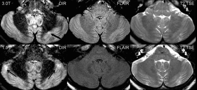
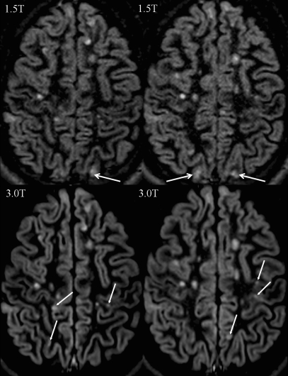
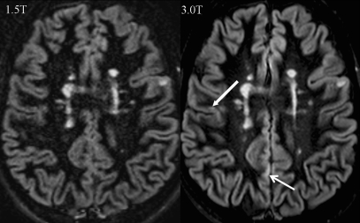
Thirty-four patients with clinically isolated syndromes or definite MS were included. All patients underwent magnetic resonance imaging (MRI) at 1.5 T and 3 T, including T2-weighted turbo spin echo (TSE), fluid-attenuated inversion recovery (FLAIR) and DIR sequences. All images were analysed for focal lesions categorised according to their anatomical location.
The total number of detected lesions was higher at 3 T across all pulse sequences. We observed significantly higher numbers of lesions involving the cortex at 3 T using a DIR sequence. DIR at 3 T showed 192% more pure intracortical (p < 0.001) and 30% more mixed grey matter-white matter lesions (p = 0.008). No significant increase in cortical lesions could be detected on the FLAIR and T2-weighted images. Using the T2-weighted and FLAIR sequences, significantly more lesions could be detected at 3 T in the infratentorial, periventricular and juxtacortical white matter.
DIR brain MR imaging at 3 T substantially improves the sensitivity of the detection of cortical lesions compared with the standard magnetic field strength of 1.5 T.
研究磁场强度为 3 特斯拉(T)对多发性硬化症(MS)患者皮质病变检出率的影响,特别是使用专用双反转恢复(DIR)脉冲序列。
纳入 34 例临床孤立综合征或明确 MS 患者。所有患者均在 1.5T 和 3T 行磁共振成像(MRI)检查,包括 T2 加权快速自旋回波(TSE)、液体衰减反转恢复(FLAIR)和 DIR 序列。所有图像均根据解剖位置进行局灶性病变分类分析。
在所有脉冲序列中,3T 检测到的病变总数均较高。使用 DIR 序列时,我们观察到 3T 时皮质病变数量显著增加。3T 下的 DIR 序列显示,单纯皮质内病变增加了 192%(p<0.001),灰质-白质混合病变增加了 30%(p=0.008)。FLAIR 和 T2 加权图像上未检测到皮质病变的显著增加。在使用 T2 加权和 FLAIR 序列时,3T 下后颅窝、脑室周围和皮质下白质的病变数量显著增加。
与标准磁场强度 1.5T 相比,3T 的 DIR 脑 MRI 可显著提高皮质病变的检出敏感性。