• 文献检索
  • 文档翻译
  • 深度研究
  • 学术资讯
  • Suppr Zotero 插件Zotero 插件
  • 邀请有礼
  • 套餐&价格
  • 历史记录
应用&插件
Suppr Zotero 插件Zotero 插件浏览器插件Mac 客户端Windows 客户端微信小程序
定价
高级版会员购买积分包购买API积分包
服务
文献检索文档翻译深度研究API 文档MCP 服务
关于我们
关于 Suppr公司介绍联系我们用户协议隐私条款
关注我们

Suppr 超能文献

核心技术专利:CN118964589B侵权必究
粤ICP备2023148730 号-1Suppr @ 2026

文献检索

告别复杂PubMed语法,用中文像聊天一样搜索,搜遍4000万医学文献。AI智能推荐,让科研检索更轻松。

立即免费搜索

文件翻译

保留排版,准确专业,支持PDF/Word/PPT等文件格式,支持 12+语言互译。

免费翻译文档

深度研究

AI帮你快速写综述,25分钟生成高质量综述,智能提取关键信息,辅助科研写作。

立即免费体验

3.0T MRI 并不改善多发性硬化症的诊断:一项多中心研究。

Three-Tesla MRI does not improve the diagnosis of multiple sclerosis: A multicenter study.

机构信息

From the Departments of Neurology (M.H.J.H., J.B., J.K.) and Radiology and Nuclear Medicine (I.D.K., M.L.d.V., M.P.W., F.B.), MS Centre Amsterdam, and Department of Epidemiology and Biostatistics (B.I.L.-W.), VU University Medical Centre; Department of Radiology and Nuclear Medicine (I.D.K.), Onze Lieve Vrouwen Gasthuis, Amsterdam, the Netherlands; Queen Square Multiple Sclerosis Centre (N.C., O.C.) and Institutes of Neurology & Healthcare Engineering (F.B.), UCL Institute of Neurology, London, UK; Department of Neurology and Psychiatry (E.S., P.P.), Sapienza University of Rome, Italy; Department of Neurology (M. Andelova, M. Amann) and Division of Neuroradiology, Department of Radiology (M. Amann, J.M.L.), University Hospital Basel; Medical Image Analysis Centre (M. Amann), Basel, Switzerland; Istituto Neurologico Mediterraneo (P.P.), Neuromed, Pozzilli (IS), Italy; Department of Neurology (C.O.-G.), Hospital Clínico San Carlos, Instituto de Investigación Sanitaria del Hospital Clínico San Carlos (IdISSC), Madrid, Spain; National Institute for Health Research (O.C., F.B.), University College London Hospitals (UCLH) Biomedical Research Centre (BRC), UK; Department of Neurosciences (C.G.), San Camillo-Forlanini Hospital, Rome, Italy; and Department of Diagnostic and Interventional Radiology and Nuclear Medicine (C.L.), St. Josef Hospital, Ruhr University, Bochum, Germany.

出版信息

Neurology. 2018 Jul 17;91(3):e249-e257. doi: 10.1212/WNL.0000000000005825. Epub 2018 Jun 20.

DOI:10.1212/WNL.0000000000005825
PMID:29925550
原文链接:https://pmc.ncbi.nlm.nih.gov/articles/PMC6059032/
Abstract

OBJECTIVE

In the work-up of patients presenting with a clinically isolated syndrome (CIS), 3T MRI might offer a higher lesion detection than 1.5T, but it remains unclear whether this affects the fulfilment of the diagnostic criteria for multiple sclerosis (MS).

METHODS

We recruited 66 patients with CIS within 6 months from symptom onset and 26 healthy controls in 6 MS centers. All participants underwent 1.5T and 3T brain and spinal cord MRI at baseline according to local optimized protocols and the MAGNIMS guidelines. Patients who had not converted to MS during follow-up received repeat brain MRI at 3-6 months and 12-15 months. The number of lesions per anatomical region was scored by 3 raters in consensus. Criteria for dissemination in space (DIS) and dissemination in time (DIT) were determined according to the 2017 revisions of the McDonald criteria.

RESULTS

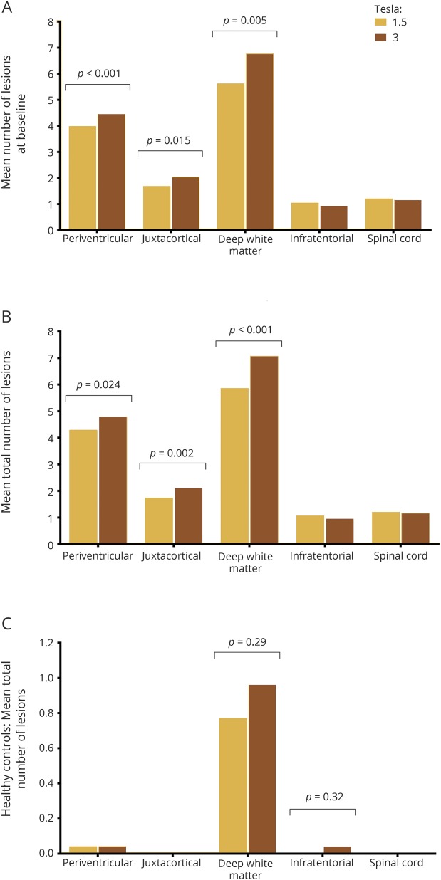
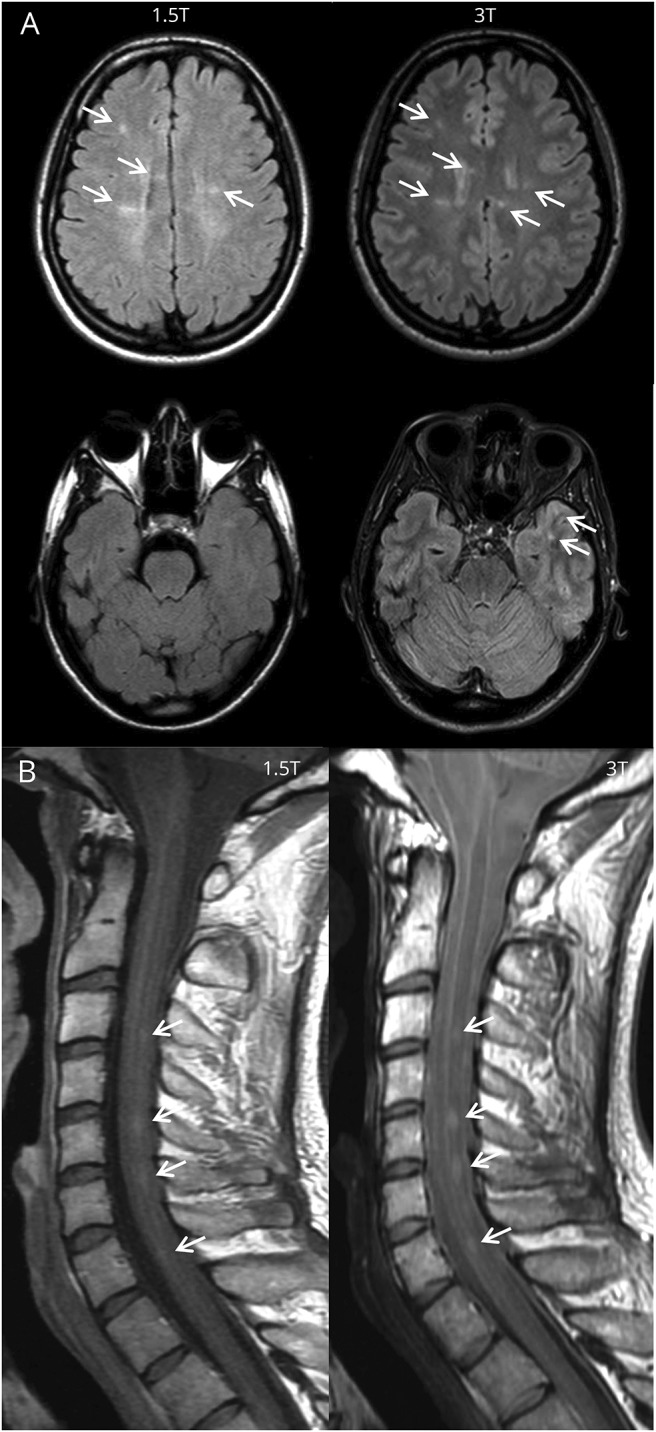
Three-Tesla MRI detected 15% more T2 brain lesions compared to 1.5T ( 0.001), which was driven by an increase in baseline detection of periventricular (12%, 0.015), (juxta)cortical (21%, 0.005), and deep white matter lesions (21%, 0.001). The detection rate of spinal cord lesions and gadolinium-enhancing lesions did not differ between field strengths. Three-Tesla MRI did not lead to a higher number of patients fulfilling the criteria for DIS or DIT, or subsequent diagnosis of MS, at any of the 3 time points.

CONCLUSION

Scanning at 3T does not influence the diagnosis of MS according to McDonald diagnostic criteria.

摘要

目的

在临床表现孤立综合征(CIS)患者的检查中,3T MRI 可能比 1.5T 检测到更多的病变,但尚不清楚这是否会影响多发性硬化症(MS)的诊断标准的满足情况。

方法

我们在 6 个 MS 中心招募了 66 名 CIS 患者(发病后 6 个月内)和 26 名健康对照者。所有参与者均根据当地优化的方案和 MAGNIMS 指南,在基线时接受 1.5T 和 3T 脑和脊髓 MRI 检查。在随访期间未转化为 MS 的患者在 3-6 个月和 12-15 个月时接受重复脑 MRI 检查。3 名评分者通过共识对每个解剖区域的病变数量进行评分。根据 2017 年 McDonald 标准修订版确定空间传播(DIS)和时间传播(DIT)标准。

结果

与 1.5T 相比,3T MRI 检测到的 T2 脑病变多 15%( 0.001),这主要是由于脑室周围(12%, 0.015)、(皮质旁)(21%, 0.005)和深部白质病变(21%, 0.001)的基线检测增加。脊髓病变和钆增强病变的检测率在不同场强之间没有差异。在任何 3 个时间点,3T MRI 均未导致更多患者符合 DIS 或 DIT 标准或随后诊断为 MS。

结论

根据 McDonald 诊断标准,3T 扫描不会影响 MS 的诊断。

https://cdn.ncbi.nlm.nih.gov/pmc/blobs/6407/6059032/a14578d86d12/NEUROLOGY2018875781FF2.jpg
https://cdn.ncbi.nlm.nih.gov/pmc/blobs/6407/6059032/4885c0b87042/NEUROLOGY2018875781FF1.jpg
https://cdn.ncbi.nlm.nih.gov/pmc/blobs/6407/6059032/a14578d86d12/NEUROLOGY2018875781FF2.jpg
https://cdn.ncbi.nlm.nih.gov/pmc/blobs/6407/6059032/4885c0b87042/NEUROLOGY2018875781FF1.jpg
https://cdn.ncbi.nlm.nih.gov/pmc/blobs/6407/6059032/a14578d86d12/NEUROLOGY2018875781FF2.jpg

相似文献

1
Three-Tesla MRI does not improve the diagnosis of multiple sclerosis: A multicenter study.3.0T MRI 并不改善多发性硬化症的诊断:一项多中心研究。
Neurology. 2018 Jul 17;91(3):e249-e257. doi: 10.1212/WNL.0000000000005825. Epub 2018 Jun 20.
2
Prediction of a multiple sclerosis diagnosis in patients with clinically isolated syndrome using the 2016 MAGNIMS and 2010 McDonald criteria: a retrospective study.采用 2016 年 MAGNIMS 和 2010 年 McDonald 标准预测临床孤立综合征患者的多发性硬化症诊断:一项回顾性研究。
Lancet Neurol. 2018 Feb;17(2):133-142. doi: 10.1016/S1474-4422(17)30469-6. Epub 2017 Dec 21.
3
Conversion of clinically isolated syndrome to multiple sclerosis: a prospective study.临床孤立综合征向多发性硬化症的转化:一项前瞻性研究。
Mult Scler Relat Disord. 2020 Sep;44:102262. doi: 10.1016/j.msard.2020.102262. Epub 2020 Jun 4.
4
MRI criteria for multiple sclerosis in patients presenting with clinically isolated syndromes: a multicentre retrospective study.临床孤立综合征患者多发性硬化的MRI标准:一项多中心回顾性研究。
Lancet Neurol. 2007 Aug;6(8):677-86. doi: 10.1016/S1474-4422(07)70176-X.
5
The role of MRI in the diagnosis of multiple sclerosis.磁共振成像在多发性硬化诊断中的作用。
Adv Neurol. 2006;98:125-46.
6
Prospective combined brain and spinal cord MRI in clinically isolated syndromes and possible early multiple sclerosis: impact on dissemination in space and time.临床孤立综合征和可能的早期多发性硬化症中脑和脊髓的前瞻性联合磁共振成像:对空间和时间播散的影响
Eur J Neurol. 2008 Dec;15(12):1359-64. doi: 10.1111/j.1468-1331.2008.02315.x.
7
Periventricular lesions and MS diagnostic criteria in young adults with typical clinically isolated syndromes.青年人有典型临床孤立综合征时,脑室周围病变与多发性硬化症诊断标准。
Mult Scler. 2017 Jun;23(7):1031-1034. doi: 10.1177/1352458516667565. Epub 2016 Sep 6.
8
Revised diagnostic criteria of multiple sclerosis.多发性硬化症的修订诊断标准。
Autoimmun Rev. 2014 Apr-May;13(4-5):518-24. doi: 10.1016/j.autrev.2014.01.012. Epub 2014 Jan 12.
9
Performance of the 2017 and 2010 Revised McDonald Criteria in Predicting MS Diagnosis After a Clinically Isolated Syndrome: A MAGNIMS Study.2017 年和 2010 年修订的 McDonald 标准在预测临床孤立综合征后 MS 诊断中的表现:MAGNIMS 研究。
Neurology. 2022 Jan 4;98(1):e1-e14. doi: 10.1212/WNL.0000000000013016. Epub 2021 Oct 29.
10
Should the symptomatic region be included in dissemination in space in MRI criteria for MS?在多发性硬化症的MRI标准中,有症状区域是否应纳入空间扩散范围?
Neurology. 2016 Aug 16;87(7):680-3. doi: 10.1212/WNL.0000000000002975. Epub 2016 Jul 15.

引用本文的文献

1
Conventional and Advanced Magnetic Resonance Imaging Biomarkers of Multiple Sclerosis in the Brain.大脑中多发性硬化症的传统和先进磁共振成像生物标志物
Cureus. 2025 Mar 2;17(3):e79914. doi: 10.7759/cureus.79914. eCollection 2025 Mar.
2
Exploring Spinal Cord Changes in Multiple Sclerosis Patients Using MRI.使用磁共振成像探索多发性硬化症患者的脊髓变化
NeuroSci. 2024 Mar 12;5(1):87-97. doi: 10.3390/neurosci5010006. eCollection 2024 Mar.
3
Scanner-specific optimisation of automated lesion segmentation in MS.基于扫描仪的 MS 自动病灶分割的优化。

本文引用的文献

1
Impact of 3 Tesla MRI on interobserver agreement in clinically isolated syndrome: A MAGNIMS multicentre study.3T MRI 对临床孤立综合征观察者间一致性的影响:MAGNIMS 多中心研究。
Mult Scler. 2019 Mar;25(3):352-360. doi: 10.1177/1352458517751647. Epub 2018 Jan 12.
2
Diagnosis of multiple sclerosis: 2017 revisions of the McDonald criteria.多发性硬化症的诊断:2017 年麦当劳标准修订版。
Lancet Neurol. 2018 Feb;17(2):162-173. doi: 10.1016/S1474-4422(17)30470-2. Epub 2017 Dec 21.
3
Brain and Spinal Cord MR Imaging Features in Multiple Sclerosis and Variants.
Neuroimage Clin. 2024;44:103680. doi: 10.1016/j.nicl.2024.103680. Epub 2024 Oct 2.
4
Detection of Spinal Cord Multiple Sclerosis Lesions Using a 3D-PSIR Sequence at 1.5 T.1.5T 下 3D-PSIR 序列检测脊髓多发性硬化病变。
Clin Neuroradiol. 2024 Jun;34(2):403-410. doi: 10.1007/s00062-023-01376-x. Epub 2024 Jan 30.
5
Prediction of the information processing speed performance in multiple sclerosis using a machine learning approach in a large multicenter magnetic resonance imaging data set.使用机器学习方法在大型多中心磁共振成像数据集预测多发性硬化症的信息处理速度表现。
Hum Brain Mapp. 2023 Jan;44(1):186-202. doi: 10.1002/hbm.26106. Epub 2022 Oct 18.
6
Sensitivity of portable low-field magnetic resonance imaging for multiple sclerosis lesions.便携式低场磁共振成像对多发性硬化病变的敏感性。
Neuroimage Clin. 2022;35:103101. doi: 10.1016/j.nicl.2022.103101. Epub 2022 Jun 27.
7
Spontaneous spinal cord infarction in Austria: a two-center comparative study.奥地利的自发性脊髓梗死:一项双中心比较研究。
Ther Adv Neurol Disord. 2022 Mar 11;15:17562864221076321. doi: 10.1177/17562864221076321. eCollection 2022.
8
Imaging of the Spinal Cord in Multiple Sclerosis: Past, Present, Future.多发性硬化症中脊髓的影像学:过去、现在、未来。
Brain Sci. 2020 Nov 13;10(11):857. doi: 10.3390/brainsci10110857.
9
Assessment of lesions on magnetic resonance imaging in multiple sclerosis: practical guidelines.多发性硬化症磁共振成像病变评估:实用指南。
Brain. 2019 Jul 1;142(7):1858-1875. doi: 10.1093/brain/awz144.
10
Advances in spinal cord imaging in multiple sclerosis.多发性硬化症脊髓成像的进展。
Ther Adv Neurol Disord. 2019 Apr 22;12:1756286419840593. doi: 10.1177/1756286419840593. eCollection 2019.
多发性硬化及其变体的脑和脊髓磁共振成像特征
Neuroimaging Clin N Am. 2017 May;27(2):205-227. doi: 10.1016/j.nic.2016.12.002.
4
Increased cortical grey matter lesion detection in multiple sclerosis with 7 T MRI: a post-mortem verification study.7T MRI 检测多发性硬化症皮质灰质病变的增加:一项死后验证研究。
Brain. 2016 May;139(Pt 5):1472-81. doi: 10.1093/brain/aww037. Epub 2016 Mar 8.
5
MRI criteria for the diagnosis of multiple sclerosis: MAGNIMS consensus guidelines.多发性硬化诊断的MRI标准:MAGNIMS共识指南。
Lancet Neurol. 2016 Mar;15(3):292-303. doi: 10.1016/S1474-4422(15)00393-2. Epub 2016 Jan 26.
6
Evidence-based guidelines: MAGNIMS consensus guidelines on the use of MRI in multiple sclerosis--establishing disease prognosis and monitoring patients.循证指南:磁共振成像在多发性硬化中的应用——建立疾病预后和监测患者的 MAGNIMS 共识指南。
Nat Rev Neurol. 2015 Oct;11(10):597-606. doi: 10.1038/nrneurol.2015.157. Epub 2015 Sep 15.
7
Ultrahigh field MRI in clinical neuroimmunology: a potential contribution to improved diagnostics and personalised disease management.临床神经免疫学中的超高场磁共振成像:对改善诊断和个性化疾病管理的潜在贡献。
EPMA J. 2015 Aug 27;6(1):16. doi: 10.1186/s13167-015-0038-y. eCollection 2015.
8
MRI in the Diagnosis and Monitoring of Multiple Sclerosis: An Update.磁共振成像在多发性硬化症诊断与监测中的应用进展
Clin Neuroradiol. 2015 Oct;25 Suppl 2:157-65. doi: 10.1007/s00062-015-0430-y. Epub 2015 Jul 23.
9
Evidence-based guidelines: MAGNIMS consensus guidelines on the use of MRI in multiple sclerosis-clinical implementation in the diagnostic process.循证指南:磁共振成像在多发性硬化诊断中的应用 MAGNIMS 共识指南——临床实施。
Nat Rev Neurol. 2015 Aug;11(8):471-82. doi: 10.1038/nrneurol.2015.106. Epub 2015 Jul 7.
10
Defining high, medium and low impact prognostic factors for developing multiple sclerosis.定义多发性硬化症发展的高、中、低影响预后因素。
Brain. 2015 Jul;138(Pt 7):1863-74. doi: 10.1093/brain/awv105. Epub 2015 Apr 21.