Division of Sleep Medicine, Department of Neurology, Beth Israel Deaconess Medical Center and Harvard Medical School, Boston, Massachusetts, United States of America.
PLoS One. 2010 Jan 20;5(1):e8788. doi: 10.1371/journal.pone.0008788.
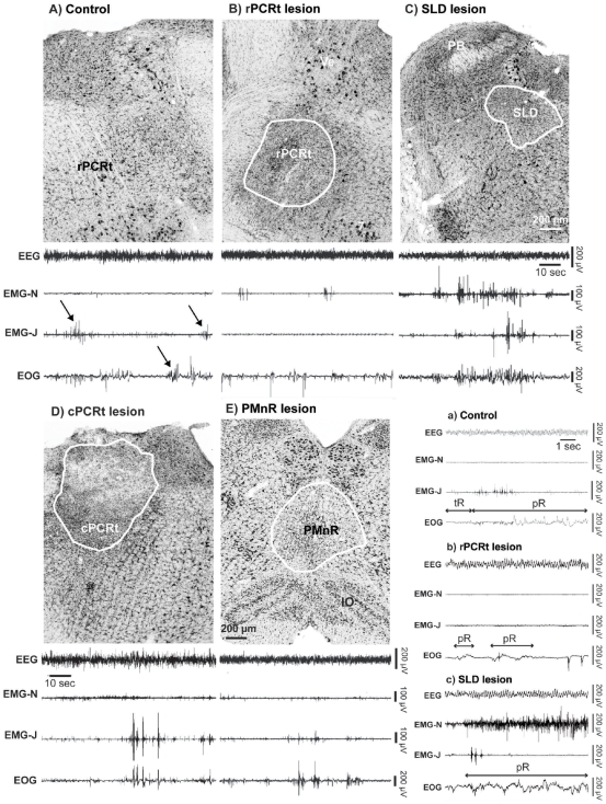
Rapid eye movement sleep (REMS) is characterized by activation of the cortical and hippocampal electroencephalogram (EEG) and atonia of non-respiratory muscles with superimposed phasic activity or twitching, particularly of cranial muscles such as those of the eye, tongue, face and jaw. While phasic activity is a characteristic feature of REMS, the neural substrates driving this activity remain unresolved. Here we investigated the neural circuits underlying masseter (jaw) phasic activity during REMS. The trigeminal motor nucleus (Mo5), which controls masseter motor function, receives glutamatergic inputs mainly from the parvocellular reticular formation (PCRt), but also from the adjacent paramedian reticular area (PMnR). On the other hand, the Mo5 and PCRt do not receive direct input from the sublaterodorsal (SLD) nucleus, a brainstem region critical for REMS atonia of postural muscles. We hypothesized that the PCRt-PMnR, but not the SLD, regulates masseter phasic activity during REMS.
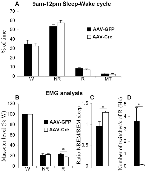
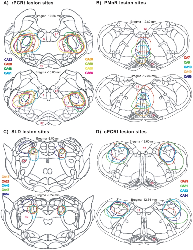
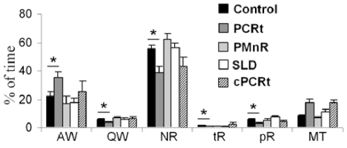
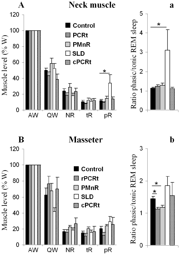
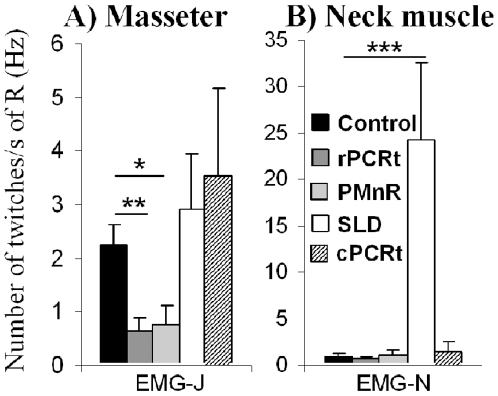
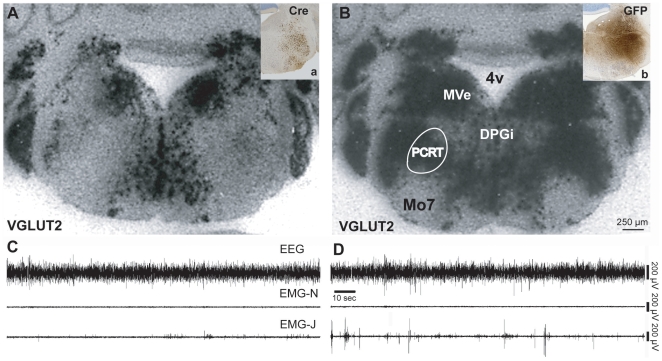
METHODOLOGY/PRINCIPAL FINDINGS: To test our hypothesis, we measured masseter electromyogram (EMG), neck muscle EMG, electrooculogram (EOG) and EEG in rats with cell-body specific lesions of the SLD, PMnR, and PCRt. Bilateral lesions of the PMnR and rostral PCRt (rPCRt), but not the caudal PCRt or SLD, reduced and eliminated REMS phasic activity of the masseter, respectively. Lesions of the PMnR and rPCRt did not, however, alter the neck EMG or EOG. To determine if rPCRt neurons use glutamate to control masseter phasic movements, we selectively blocked glutamate release by rPCRt neurons using a Cre-lox mouse system. Genetic disruption of glutamate neurotransmission by rPCRt neurons blocked masseter phasic activity during REMS.
CONCLUSIONS/SIGNIFICANCE: These results indicate that (1) premotor glutamatergic neurons in the medullary rPCRt and PMnR are involved in generating phasic activity in the masseter muscles, but not phasic eye movements, during REMS; and (2) separate brainstem neural circuits control postural and cranial muscle phasic activity during REMS.
快速眼动睡眠 (REMS) 的特征是皮质和海马脑电图 (EEG) 的激活以及非呼吸肌的弛缓,同时伴有相位活动或抽搐,特别是颅部肌肉,如眼、舌、面和颌部的肌肉。虽然相位活动是 REMS 的一个特征,但驱动这种活动的神经基质仍未解决。在这里,我们研究了 REMS 期间咀嚼肌(颌部)相位活动的神经回路。控制咀嚼肌运动功能的三叉神经运动核 (Mo5) 主要接收来自小细胞网状结构 (PCRt) 的谷氨酸能输入,但也接收来自相邻旁正中网状区域 (PMnR) 的输入。另一方面,Mo5 和 PCRt 没有直接接收来自脑干中对于姿势肌 REMS 弛缓至关重要的 sublaterodorsal (SLD) 核的输入。我们假设 PCRt-PMnR,但不是 SLD,调节 REMS 期间咀嚼肌的相位活动。
方法/主要发现:为了验证我们的假设,我们在 SLD、PMnR 和 PCRt 的细胞体特异性损伤的大鼠中测量了咀嚼肌肌电图 (EMG)、颈部肌肉 EMG、眼电图 (EOG) 和 EEG。双侧 PMnR 和头侧 PCRt(rPCRt)损伤分别减少和消除了 REMS 咀嚼肌的相位活动,但尾侧 PCRt 或 SLD 损伤没有改变颈部 EMG 或 EOG。为了确定 rPCRt 神经元是否使用谷氨酸来控制咀嚼肌的相位运动,我们使用 Cre-lox 小鼠系统选择性地阻断 rPCRt 神经元的谷氨酸释放。rPCRt 神经元的谷氨酸神经传递的遗传破坏阻断了 REMS 期间咀嚼肌的相位活动。
结论/意义:这些结果表明:(1) 延髓 rPCRt 和 PMnR 中的运动前谷氨酸能神经元参与了 REMS 期间咀嚼肌相位活动的产生,但不参与相位眼运动;(2) 不同的脑干神经回路控制 REMS 期间姿势肌和颅肌的相位活动。